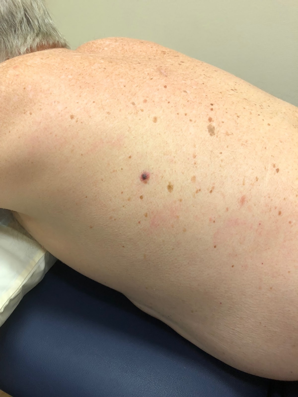
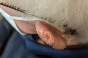
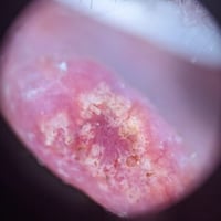
Case discussion: How would you treat this patient? [22 May]

In this week’s case discussion from Dr David Stewart, we look at a 84-year-old male patient who was seen by another GP with a new tender lesion on the rim of his helix. That GP gave him antibiotics but the lesion didn’t improve.

The patient has a history of squamous cell carcinoma and lentigo maligna.

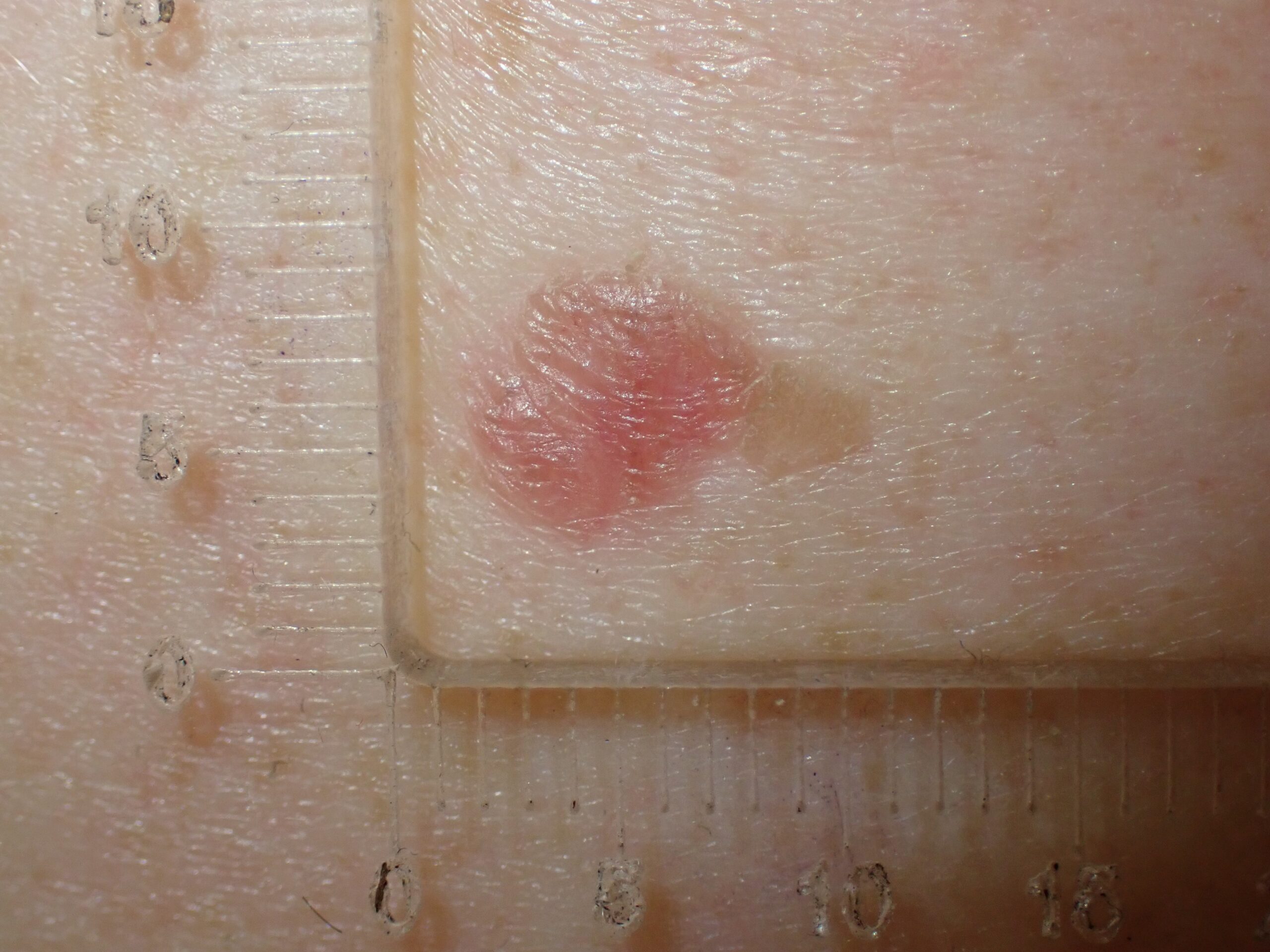
What do you think of this lesion? What would you do next?

case discussion

Update

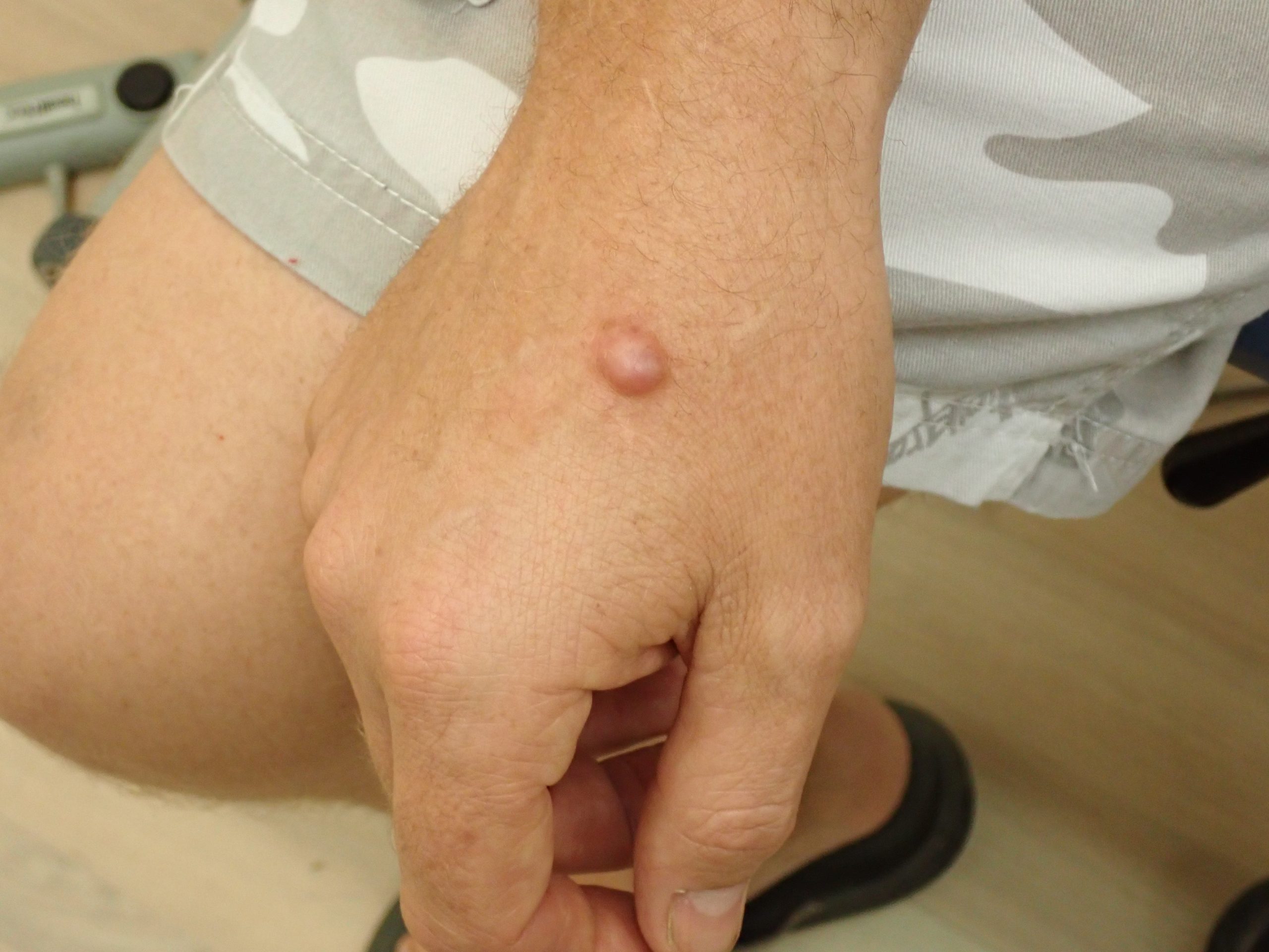
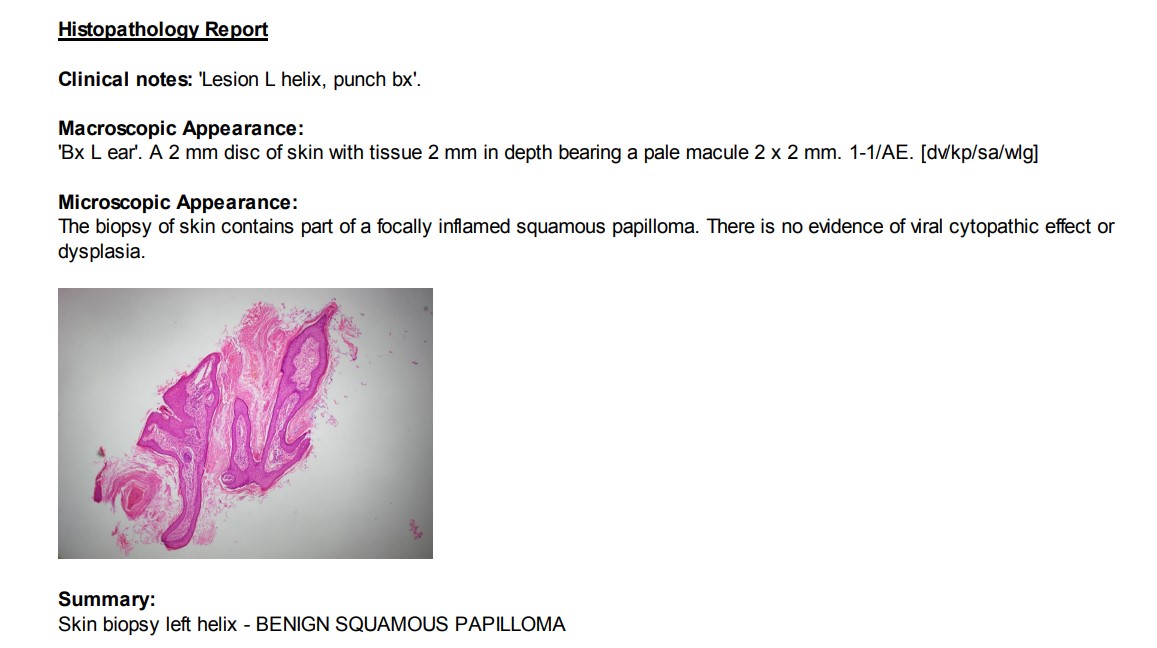
Here is the pathology result. As the management of benign lesions is important, too, what would you do next?


For further information on this topic, you may be interested to learn more about the HealthCert Professional Diploma program in Skin Cancer Medicine.

case discussion


Would you like to obtain advice or share your experience with your colleagues in the weekly blog case discussion, moderated by Dr Terry Harvey?

 Participate with your cases so that we can learn together!

Submit your case here or send details to [email protected]


MORE CASE DISCUSSIONS

Leave a Reply

Your email address will not be published. Required fields are marked *

26 comments on “Case discussion: How would you treat this patient? [22 May]

      1. Hi Terry,
        Interesting to read the results of benign papillomatous lesion, which would not expect to see arborizing vessels. Here I will do curettage or shave biopsy for the whole lesion.
        What are your thoughts about this?
        Regards
        Randa

  1. punch biopsy for ?scc. decide excision involved cartilage or not. Helical double advancement flap for repair after excision.

  2. Chondrodermatitis nodularies helices.. can try steroids.. if not help must need biopsy as H/O SCc

  3. Non pigmented lesion
    Prediction without pigment applies
    There is presence of ulcer and white clues including white circles as well as surface keratin.(Vessels are not clear)
    Clinically squamous cell carcinoma.
    I would do a biopsy to confirm the diagnosis
    The definitive treatment is excision with 2mm margin ,closure option is an advancement helical flap.

  4. Pink helical lesion with abnormal vasculature on surrounding ‘chaotic’ rim
    Suspicious for SCC DDx Chondrodermatitis nodularis chronica helicis
    For punch biopsy and if cancer confirmed for wedge excision

  5. Clinically a NMSC until proved otherwise. A tender keratotic lesion with a central necrotic ulcer.
    Dermatoscopic image= A keratotic rim with a central erythematous crater? ulcer, with some white lines and abnormal BVs. The keratotic rim has white circles and radial linear irregular BVs.
    IMPRESSION = more likely to be a SCC.
    PLAN= A 3 mm punch biopsy at the rim and center. To determine the histological diagnosis and degree of invasion? PIN involvement etc.

  6. Appearances are suspicious for a SCC. I would do a punch biopsy to get a diagnosis and then either a double advancement flap or a wedge resection to excise the lesion if biopsy proved an SCC.

  7. Scaly and tender with a suggestion of white circles is suspicious for IESCC/SCC. I would do a 3-4mm punch biopsy and if the diagnosis is confirmed, an H-flap. Chondrodermatitis is possible, but less likely on the top of the ear where there is less pressure.

  8. tender, indurated, keratotic lesion, likely SCC; I would do a punch Bx to confirm and to get info re differentiation, PNI/LVI and then will need to be excised if malignancy confirmed

  9. 84 yo should have a big ear to play with
    Definitely wedge excision and repair under periauricular block
    Pathology should be SCC or less likely traumatised inflamed Chondrodermatitis Helicus

  10. Chondrodermatitis nodularis helicis presents as a raised nodule on the parts of the ear cartilage referred to as the “helix” or the “antihelix.” These are the inner and outer ridges of cartilage in the ear.
    Management includes, excision, cryotherapy. Laser or take pressure off the lesion when sleeping

    1. Hi Everyone

      A few people were aiming for large and complex excision plans as a first step.

      Whilst for obvious BCCs and SCCs in simple areas I will often offer curative treatment as the first procedure – it would be extremely rare for me to do a complex procedure on the head/neck without being 100% sure it is required. A biopsy before definitive treatment in my view is a must if a complex procedure is planned, or if the lesion is on the face.

      There is legal precedent within Australia where a doctor was successfully sued for performing a flap repair on the nose and the result returned as an inflamed hair follicle, not the BCC that they were expecting.

  11. Hi Everyone

    A few people were aiming for large and complex excision plans as a first step.

    Whilst for obvious BCCs and SCCs in simple areas I will often offer curative treatment as the first procedure – it would be extremely rare for me to do a complex procedure on the head/neck without being 100% sure it is required. A biopsy before definitive treatment in my view is a must if a complex procedure is planned, or if the lesion is on the face.

    There is legal precedent within Australia where a doctor was successfully sued for performing a flap repair on the nose and the result returned as an inflamed hair follicle, not the BCC that they were expecting.

  12. The biopsy, only a 4mm piece, is benign. However there is a history of SCC and lentigo maligna.
    If conservative therapy with further course of antibiotics and topical steroid failures to heal, then consider wedge resection to eliminate the lesion and subject it all to histological examination.