#=$visible?>
Case discussion: How would you treat this patient? [15 May]
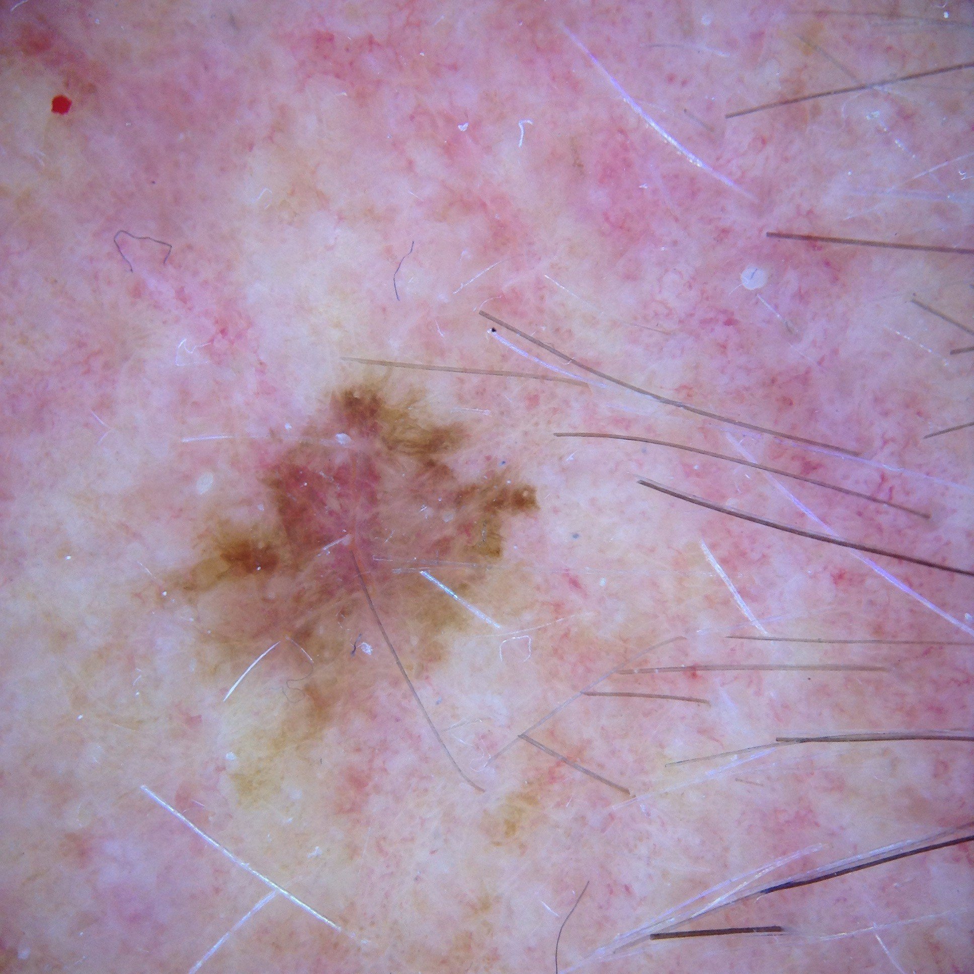
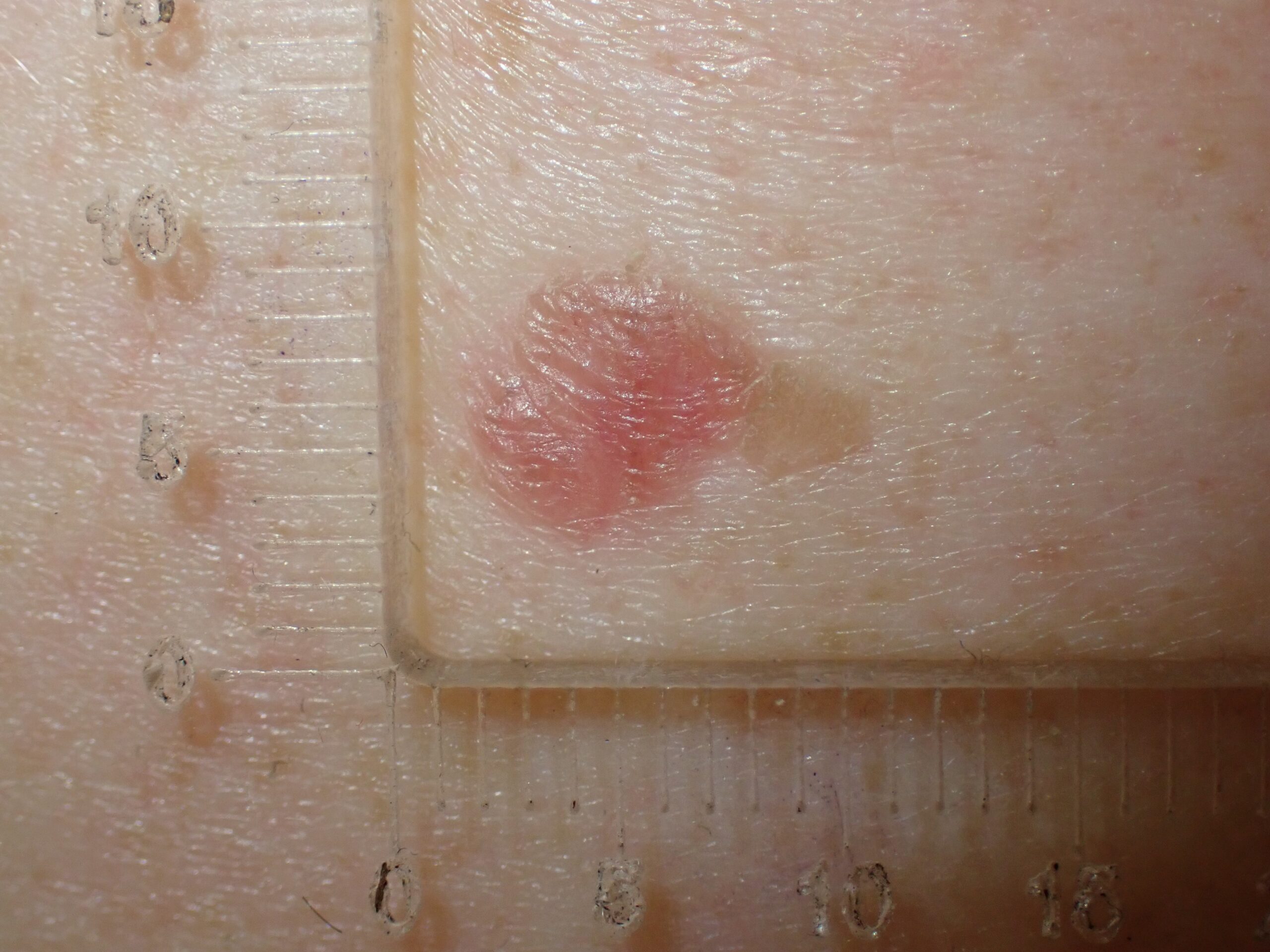
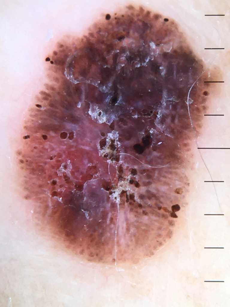
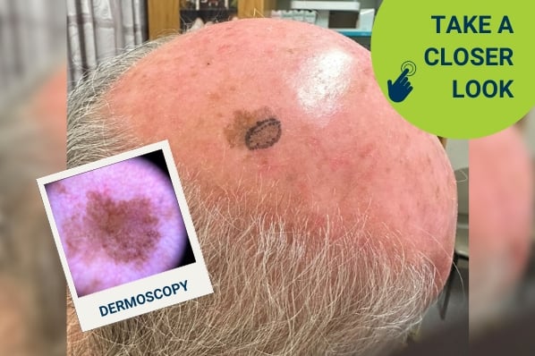
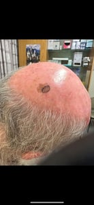
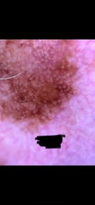
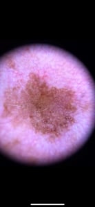
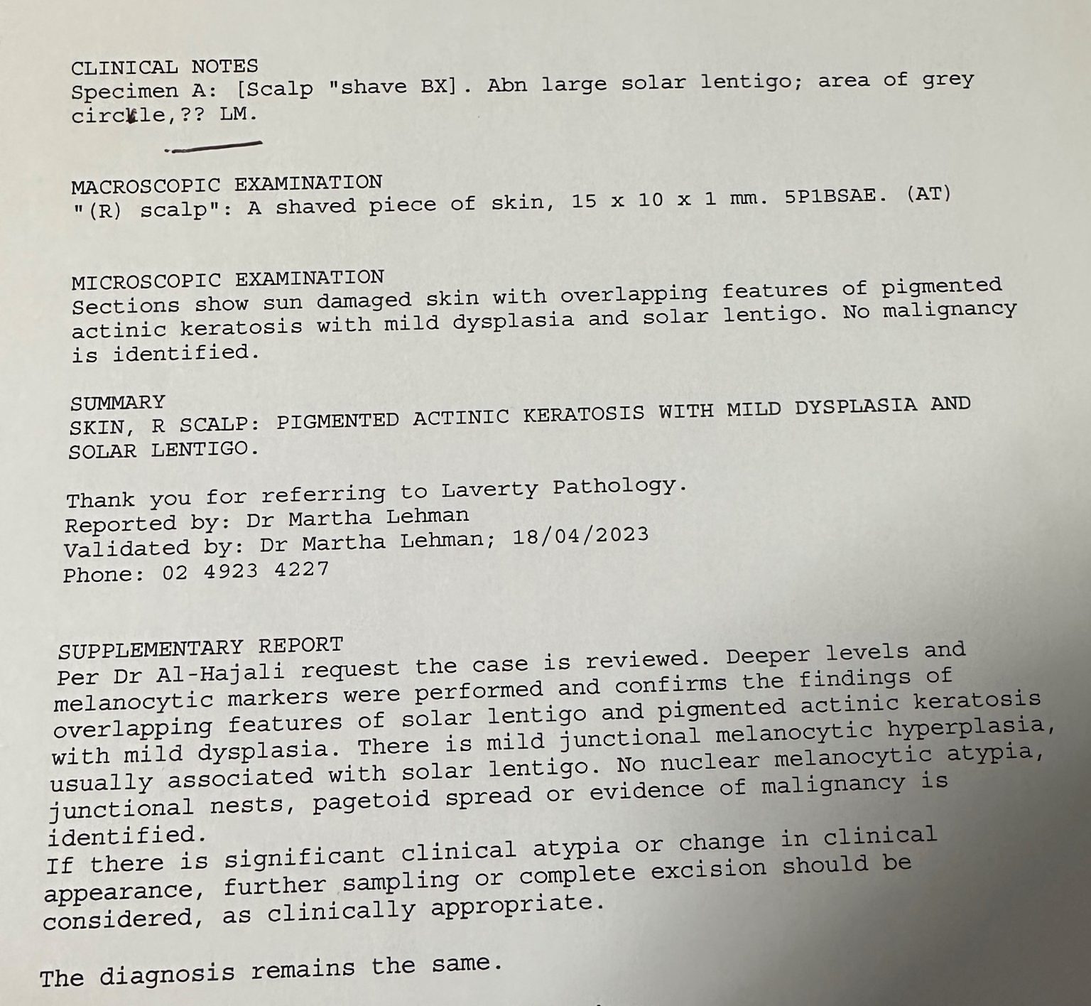
In this week’s case discussion, submitted by Dr Randa Al-Hajali, we look at a 76-year-old male patient who presented for his routine skin check. This large patch was noted on the right parietal scalp.
What do you think of this lesion? What would you do next?
Update
For further information on this topic, you may be interested to learn more about the HealthCert Professional Diploma program in Skin Cancer Medicine.
Would you like to obtain advice or share your experience with your colleagues in the weekly blog case discussion, moderated by Dr Terry Harvey?
Participate with your cases so that we can learn together!
Submit your case here or send details to [email protected]
Leave a Reply Cancel reply
20 comments on “Case discussion: How would you treat this patient? [15 May]”
Subscribe
Receive the newest case studies, free video tutorials and research articles right in your inbox.
Lentigo Maligna
Shave biopsy
Would take a thorough Hx about this lesion
Possibility of lentigi maligna and shave / excision biopsy . He may need a referral to plastics/dermatologist given the size
Melanoma
This has features of melanoma-in-situ. I would take a large shave of the 5 o’clock .
Large brown macule on his scalp Dermatoscopic image = lentigo maligna vs a lentigo. A non-polaroid image would have been good to look at the surface follicles/blue structures.
Lentigo maligna until proved otherwise re atypical pigmented follicular openings. Grey annular granular follicles, Brown pigmented asymmetrical follicle openings. Double circles and obliteration of a follicular opening. Odd, angulated line?
Lentigo features, lentiginous network, sharp mouth eaten boarders + variable follicular openings.
IMPRESSION = lentigo maligna/lentignous melanoma until proved otherwise
PLAN= a shave biopsy.
I am an MBBS, passed Diploma Dermatology from Australia & part Fellowship from National Skin Center, Singapore.
This is Melanoma in Situ
Assymetry
Grey colour
Perifollicular pigmentation ,assymetric
Annular granular pattern
Management Excision biopsy with 2-3 mm margins
Sometimes Pigmented Seb K with Lichenificantion could be a differential but given his age I would err on side of caution.
dermoscopy not very clear but macroscopically appears to be a pigmented actinic keratosis grade 2
Pigmented Lesion. Asymmetrical in pattern and colour. Thicker darker lines over suspicious area and pigmented circles. Suspicious for change to Lentigo Maligna Melanoma. Shave excisional biopsy of the entire lesion with 2mm margin clearance
Hi Everyone
The classic trio of flat pigmented lesions on the head and neck are:
– flat lentigo/seborrheic keratosis
– pigmented actinic keratosis
– lentigo maligna
And also combinations of these three…
Place your bets!
I agree but one has to be concerned to rule out melanoma .
Although I do not usually concur with excision before a definitive diagnosis , this may be an exception.
However a physical examination would provide better confidence.
Then however , this is a good case for confocal microscopic examination
Thanks, Tom.
I agree in that the impacts and risks of surgery/sampling always have to be balanced against the possibility of malignancy.
I only offer diagnostic sampling when I believe the risk of the procedure is lower than the risk of the lesion.
“If in doubt, cut it out” sounds clever because it rhymes, but if we practice medicine like that then every child with a cough would get a high resolution CT to look for lung cancer.
There are at times other ways to gain more information without surgery – this may be monitoring the lesion where it is safe to do so, or confocal microscopy
Multiple colours including grey, circle within a circle, asymmetrical pigmentation follicular openings, looks like lentigo maligna; I would do 2-3 punch biopsies of the most abnormal areas which are at 4-8 o’clock on the larger/2nd dermoscopic image
MELANOMA
Looks like a superficial melanoma
Would want to rule out it not being a pigmented solar keratosis
shave biopsy sample for histology
lentigo maligna
I would take biopsies of the most suspicious looking areas
chaos: asymmetrical structures, colours; indistinct borders, multiple colours, diameter >5mm,
clues: grey circles face/scalp
DDx: differential diagnosis Lentigo maligna
Rx: excisional shave biopsy
A broad shave sample of a flat pigmented lesion on the head/neck where there is no evidence of a melanocytic proliferation is very strong evidence that this is not a lentigo maligna despite the fact that the whole lesion hasn’t been submitted to pathology.
What would be your follow-up plan for this lesion/patient?
I arranged a follow up after 3 months.
I was also surprised at the pathology results since my clinical diagnosis was strong for LM.
Collided lesion like this is very confusing with melanoma. Regards
Randa
I feel it is a lentigo malignant melanoma – it has those thicker follicular borders in the area sampled.
Surprised at the pathology but I guess that is the gold standard