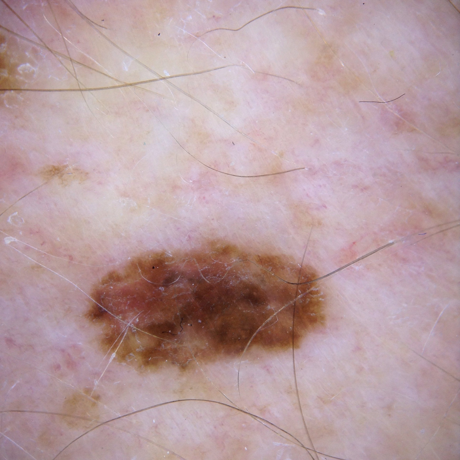
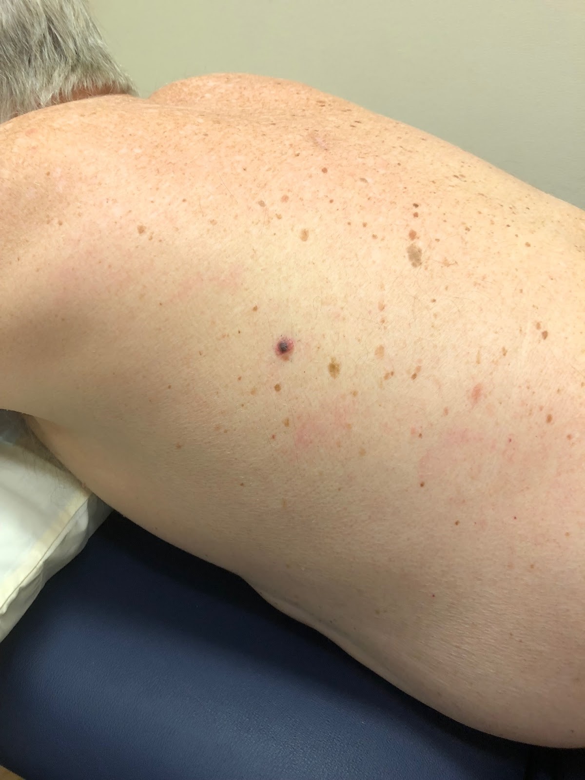
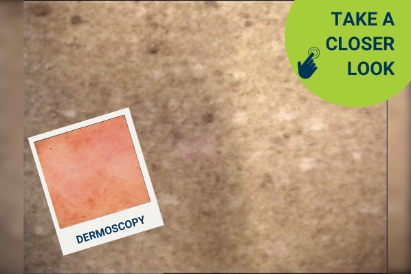
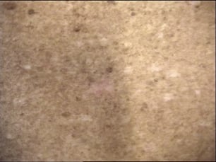
#=$visible?>
Case discussion: How would you treat this patient? [8 May]
In this week’s case discussion from Dr Peter Ie, we look at a 58-year-old male patient with a non-pigmented, pink, smooth lesion found on the lower back during a routine skin cancer check.
What are the possible differential diagnoses? What is your plan for follow-up?
Update
For further information on this topic, you may be interested to learn more about the HealthCert Professional Diploma program in Skin Cancer Medicine.
Would you like to obtain advice or share your experience with your colleagues in the weekly blog case discussion, moderated by Dr Terry Harvey?
Participate with your cases so that we can learn together!
Submit your case here or send details to [email protected]
Leave a Reply Cancel reply
11 comments on “Case discussion: How would you treat this patient? [8 May]”
Subscribe
Receive the newest case studies, free video tutorials and research articles right in your inbox.
Punch biopsy and follow up appointment
Looks sBCC
Check the patient if Benign after 6 months for a follow-up visit doctor. If suspicious need Biopsy.
I have a patient in early 70s with multiple areas similar appearance for 10 years. Treated multiple times by other GP as superficial BCC with cryotherapy.Biopsy was done previously that had shown BCC. Do we biopsy again from few sites???
I see a lot of these, though usually the vessels are easier to see. I suspect them to be superficial BCCS and give them double cryo.
White lines and structureless lesion. Smooth and no scale making keratitis lesion unlikely . Can’t tell the size . White lines can be seen in BCC and melanoma apart from other diagnoses . I would saucerize shave at an adequately deep level and 2-3mm beyond obvious border as can’t afford to wait for change. This will give the diagnosis without delay and manage accordingly . Punch biopsy risks incorrect histo assessment as may be regression
“Can’t afford to wait for change” is a great comment.
There are certainly times when it is perfectly safe to watch a lesion over time, and times when that is not a good idea.
If melanoma is part of your differential and the lesion is raised, then the ‘clock is already ticking’ as it is potentially invasive melanoma and a life-threatening lesion. You cannot afford to wait for it to change.
If a naevus is flat with no signs of deep melanoma, it can be safe to watch it over a short period of three months to try and avoid an unnecessary excision.
If the differential is only between BCC and lichenoid keratosis – it is perfectly safe to watch it for a few months and see how it evolves.
This white lesion is different from the others on the back. Larger, very irregular, crystalline structures, and white areas would could indicate a lesion comprising mainly of regression. It is suspicious.
Would shave biopsy / excision and take it from there.
Stands out as a pink/white structureless area amongst multiple lentigo and sun damaged skin. Suspicious for something. Superficial BCC would be the probability diagnosis. Shave biopsy would be appropriate.
It has a lot of white suggesting abnormal collagen production. ??morphoeic bcc or some kind of desmoplastic melanoma..??
How long has it been there and how big is it..?
It is sitting in a field of sun damaged skin so a malignancy needs to be considered. Do a punch biopsy or if little take it out.
possible diagnosis is ?bcc ?scc ?dermatofibroma,
I would suggest shave biopsy to confirm the diagnosis.