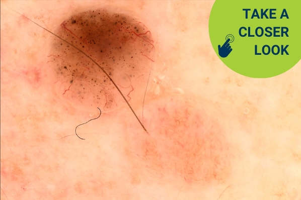
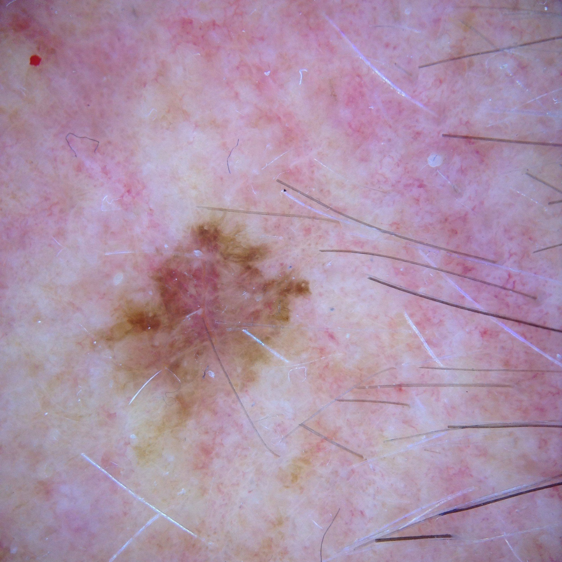
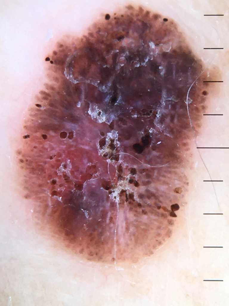
Case discussion: How would you treat this patient? [24 April]

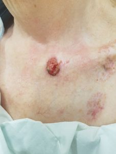
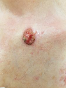
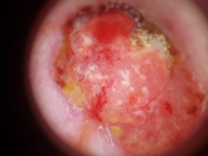
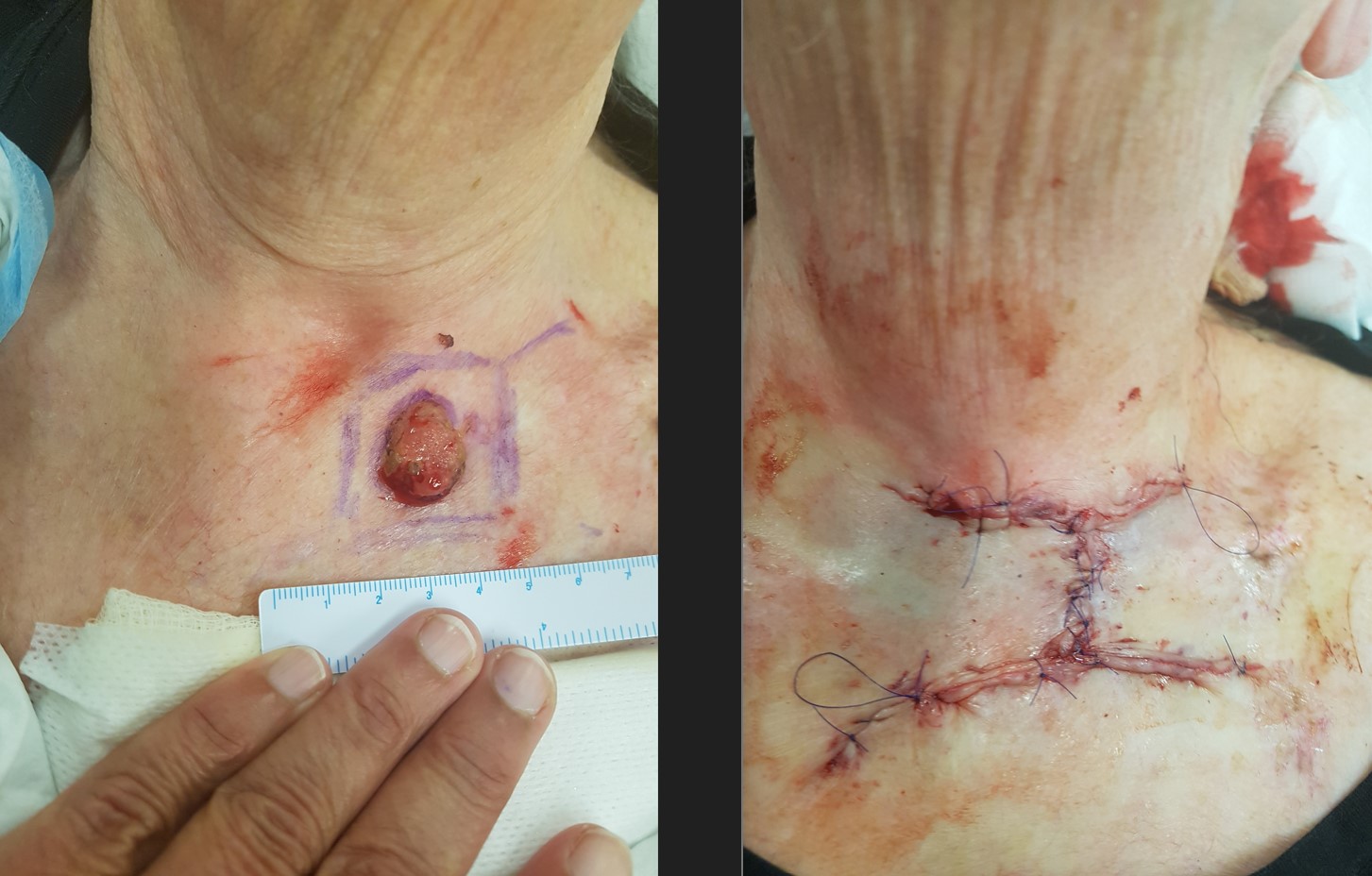
In this week’s case discussion, submitted by Dr Renuka Ranasinghe, we look at a 76-year-old female patient who presented with a large ulcerating area on her neck.

What is your diagnosis, and how would you manage this lesion?

case discussion

Update

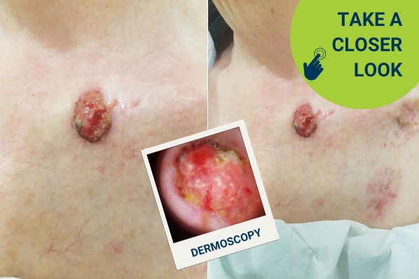
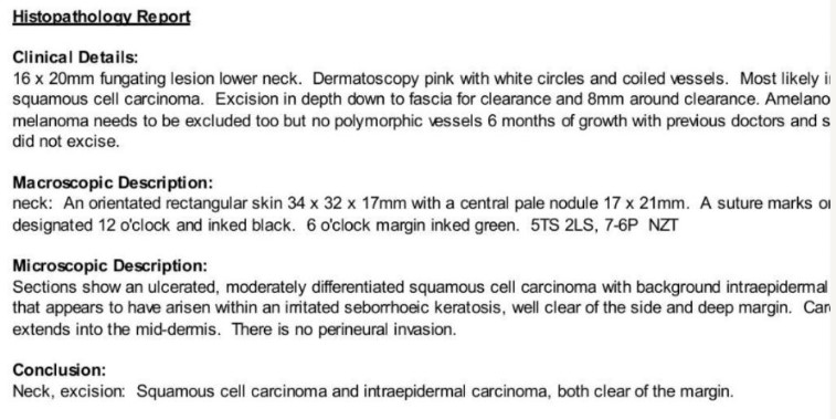
Here is the histopathology report (apologies for the cut-off edge), plus surgery photos.


For further information on this topic, you may be interested to learn more about the HealthCert Professional Diploma program in Skin Cancer Medicine.

case discussion


Would you like to obtain advice or share your experience with your colleagues in the weekly blog case discussion, moderated by Dr Terry Harvey?

 Participate with your cases so that we can learn together!

Submit your case here or send details to [email protected]


MORE CASE DISCUSSIONS

Leave a Reply

Your email address will not be published. Required fields are marked *

21 comments on “Case discussion: How would you treat this patient? [24 April]

  1. Ulcerated red nodule: malignant.
    Biopsy or immediate excision. My prov dx would be poorly differentiated SCC

  2. Appearances strongly suggest malignancy. Possibly BCC.
    For punch biopsy then excision according to histology.

  3. Would like to know more history. I’ve seen similar before -usually SCC but ulcerated BCCs can look like this.

    1. Hi Peter

      History is important in some cases – but how would it alter your plan here?

      If the patient said it had always been there – would you leave this lesion alone with no follow up?

  4. Esophageal melanoma usually presents with progressively worsening dysphagia, and patients often present late in the disease course. Esophageal melanoma can be treated with surgical resection, chemotherapy, targeted therapy, or immunotherapy depending on the stage and tumor mutations. However, due to the rarity of the disease, no trials have been performed to deliver a gold standard of treatment. We present the case of a 76-year-old female who was diagnosed with metastatic primary malignant melanoma of the esophagus and underwent treatment with nivolumab, a PD-1 receptor antagonist.

    Keywords: primary malignant melanoma, dysphagia, nivolumab, melanoma, esophageal melanoma

  5. Nasty lesion – could be a amelanotic melanoma! I am unable to see too many polymorphic vessels but higher diff scc is did. excision by with 2 mmm margins preferred urgently or even a punch on the day to exclud mm

  6. This looks like a BCC, but definitely needs excision. It seems to have a large horizontal pale surrounding that may define the edge. I use my nail to find the edge, but also a punch biopsy often makes the edge more obvious. If I was certain of the boundary I would excise it (probably horizontally, depending on skin laxity) with a 2mm margin. This lesion is quite advanced and may need radiotherapy, depending on the pathology. I’m also concerned about the 2 pigmented lesion on the right, that may also need biopsy.

    1. Heather the pigmentation around is atrophy and inflammation from Radiotherapy she had over 20 years ago and cannot remember.

  7. appears to be a moderately differentiated SCC likely or a poorly differentiated one, needs a punch done 4mm for diagnosis and then aim a flap

    1. Hi Ashwin

      Is there any reason you would aim for a flap rather than primary closure?

      The aim should always be the simplest procedure that treats the lesion.

      1. Hi Terry her upper chest skin was so thin and atrophic , the defect post excision was too large for primary closure. Thanks for posting my cases

  8. Raised , popular . Rolled up edges, bleeding, at places necrotic tissue
    My first differential diagnosis will be SCC
    Second possibility is modular hypertrophic BCC

  9. Clinically this does not look good. This is a non-pigmented nodular ulcerated vascular lesion.
    Dermatoscopic image = an ulcerated raised vascular lesion with some red/cherry red clods/as well as structureless red/cherry red areas. The clods have some linear irregular centered blood vessels. (This is not normal) Some exudate at the margin.
    IMPRESSION= an amelanotic nodular melanoma vs a merkel cell carcinoma vs a poorly differentiated SCC vs an atypical fibroxanthoma
    PLAN = urgent excision biopsy

    1. I agree with this assessment and would also add a possibility of a cutaneous metastasis – we don’t know anything about the medical history of this elderly lady.

  10. A nodular ulcerated vascular lesion as well as some red/cherry red areas.Malignancy until proven otherwise– therefore excision ,with consideration to include the possible extension in the 2 o’clock position.