Case discussion: How would you treat this patient? [20 February]

In this week’s discussion, submitted by Dr Renuka Ranasinghe, we look at the challenging case of an 88-year-old lady with Dementia and Parkinson’s Disease who came in for a skin check. She is wheelchair bound, and frequent writhing movements were an issue during examination and treatment (therefore, apologies for the blur in the photos).
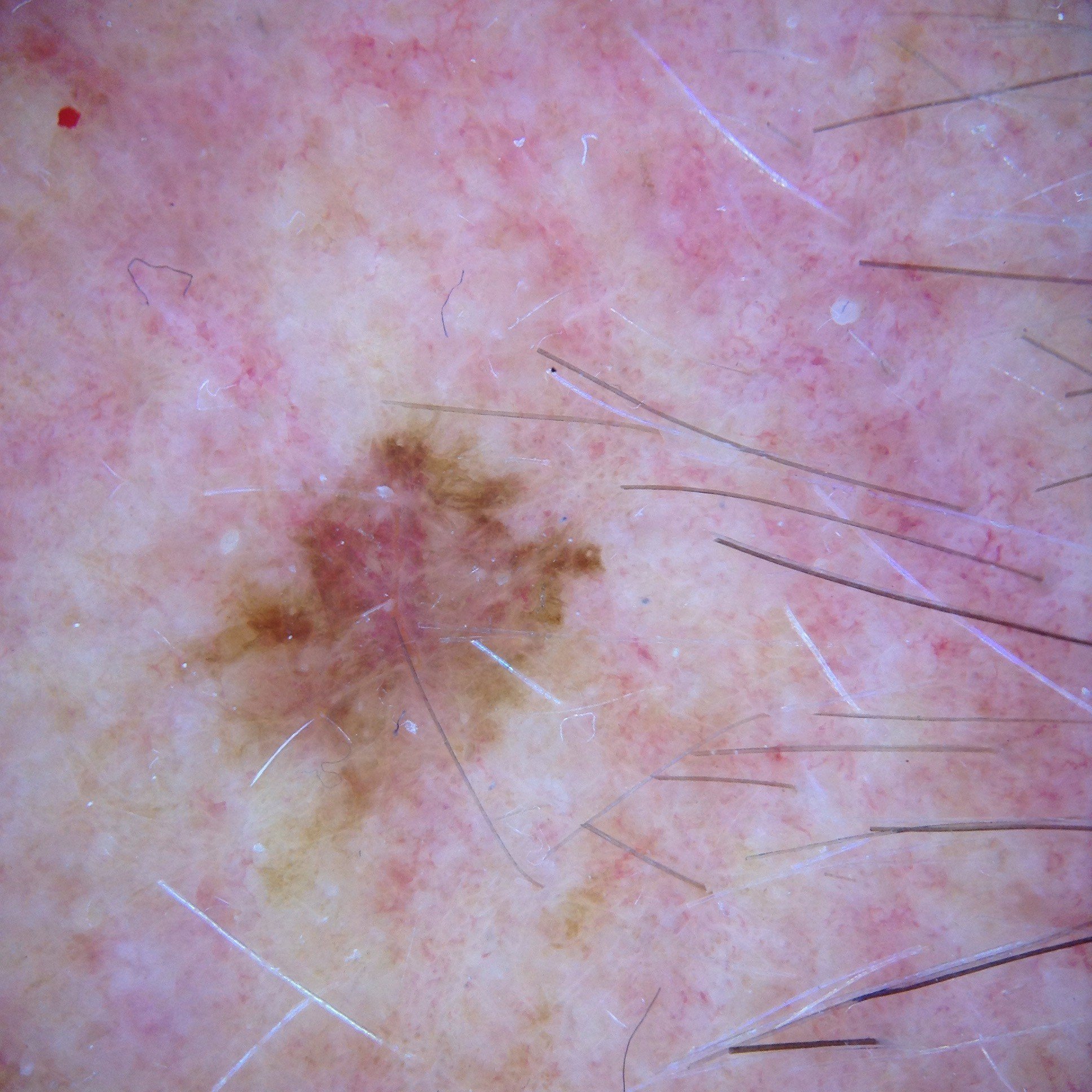
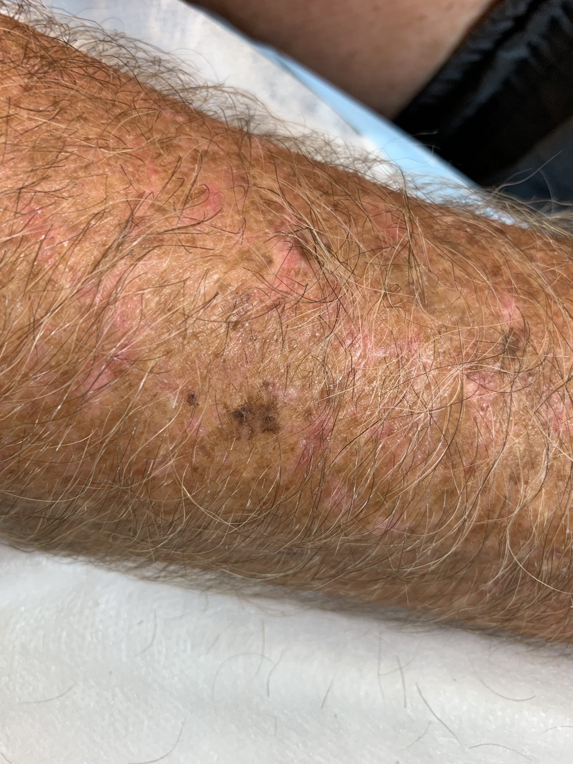
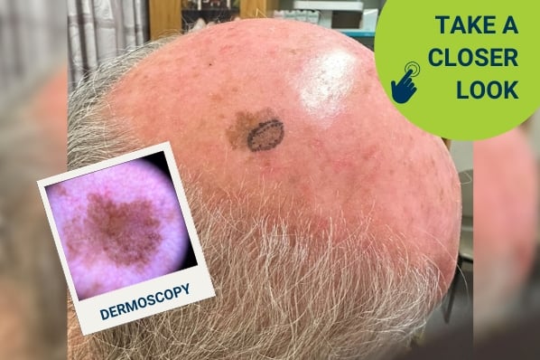
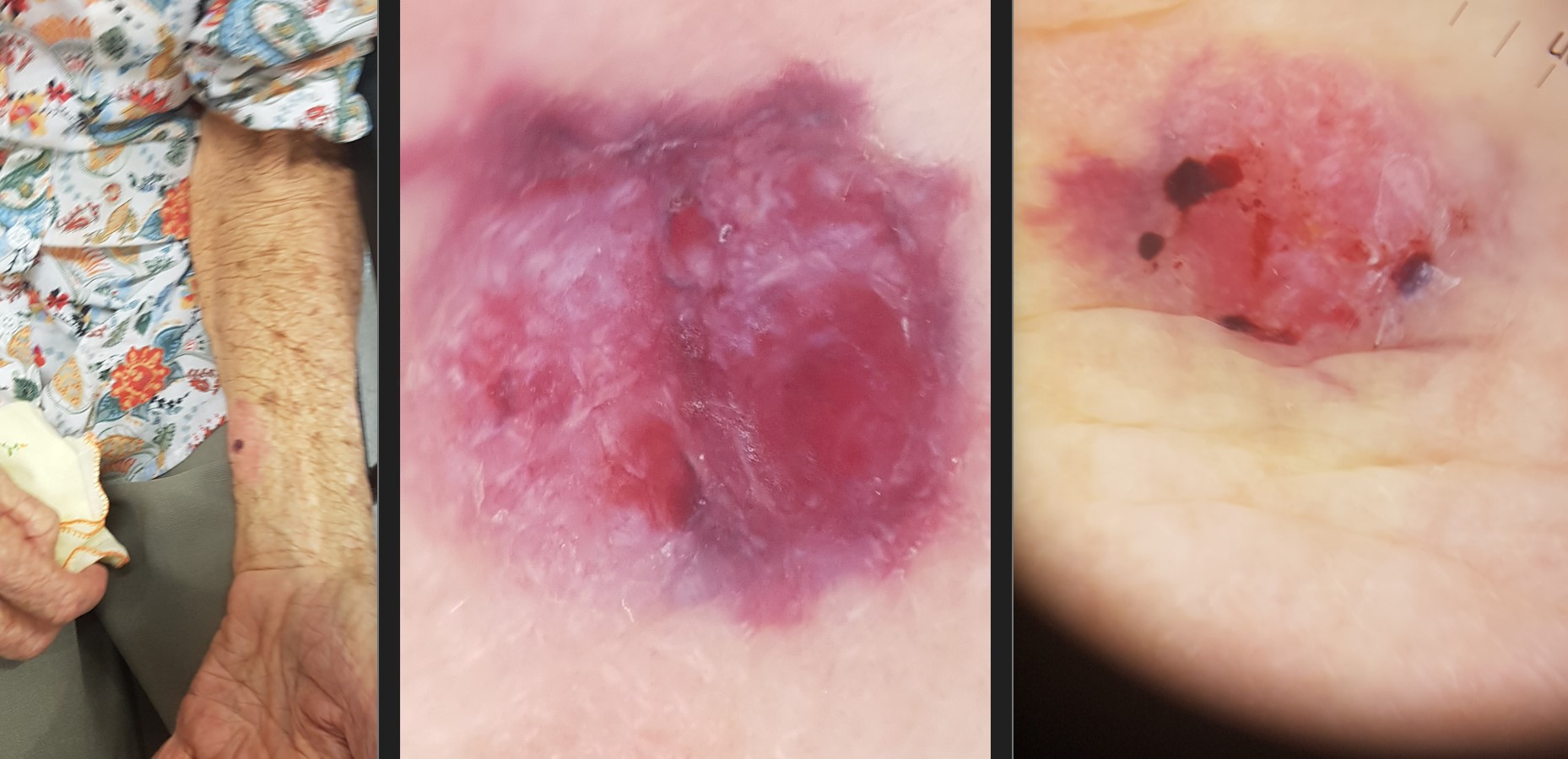
According to the patient’s daughter, a pink nodular lesion on the patient’s arm had remained unchanged for years but had shown noticeable change over the past few weeks. The photos below show the dermoscopic change that occurred between the patient’s initial skin exam and a follow-up check two weeks later.

What are your thoughts here? With consideration of the patient’s other conditions, how would you manage this case?

Update:

Here is the pathology report:
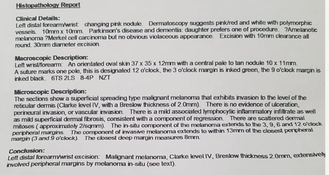
case discussion

The patient’s family wanted one-off surgery due to her clinical condition.

The invasive melanoma was fully excised with 13mm closest peripheral margin and 8mm deep margin clearance. Closed with rhomboid flap from volar aspect of forearm.

Melanoma in situ extends to the peripheral margins. Referred to MDT team in Perth and awaiting advice.

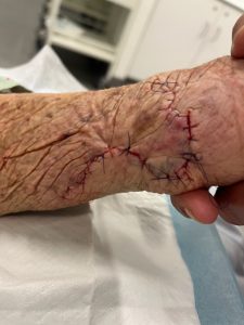
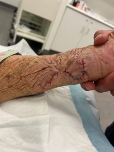
Post-surgery photos:

case discussion

For further information on this topic, you may be interested to learn more about the HealthCert Professional Diploma program in Skin Cancer Medicine.


case discussion


Would you like to obtain advice or share your experience with your colleagues in the weekly blog case discussion?

 Participate with your cases so that we can learn together!

Submit your case here or send details to [email protected]


MORE CASE DISCUSSIONS


Leave a Reply

Your email address will not be published. Required fields are marked *

15 comments on “Case discussion: How would you treat this patient? [20 February]

  1. Tricky real world case. Given age and comorbidities, the usual “excision biopsy and go from there” may not be best option (difficult excision, high risk of wound complications given dementia), but the dermoscopy looks potentially alarming so just “giving it a freeze” also not really an option. Part of management is discussions with family, but I’d be inclined to do a quick shave biopsy just to get a diagnosis – it’s not ideal for nodular lesions but in this patient it would be quick and simple, and even if it comes back as melanoma its not the end of the world if we don’t get a full thickness sample as the option of surgery may not be the way to go. If it comes back as a SCC or BCC, then maybe malignant cryo might be tolerated. I’ll be interested in other peoples thoughts though – good case!

  2. Pink white nodular lesion; elderly patient sun damaged skin; rapidly growing.
    Likely amelanocitic melanoma
    Excision biopsy 2mm margins

    DDx SCC

  3. Marginal pigment network, milky pink veil, atypical vessels, given age of patient Amelanotic Melanoma would be first on list. Excision with 2 mm margins

  4. Hi, my first option is Amelanotic Melanoma . Is very important the chrysalis or perpendicular white lines. I think the best is an excicional Biopsy 2,5cm Margin)

  5. ? Amelanotic melanoma – 88yo with dementia and Parkinson’s. I would discuss with family but probably advise to leave untreated

  6. The lesion appears to have grown in a surroundings in red area in which there appears to be the complete absence of pigment. Dermoscopally There seems to be a milky pink colour with short, white lines and some ulceration. The vessels are not clear enough to be able to describe. Its rapid growth and complete lack of pigment suggests a non-melanoma malignancy , although amelanotic melanoma is not excluded on that because the history may be in accurate. It raises the possibility of a poorly differentiated, SCC, without much scale, mierkels or even AFX. I personally I would put in some local and scoop it out in a decent way with a seven mm curette as deeply as I could and send it off to see what it was. Possible further curating and diathermy and leave it alone afterwards would be strongly indicated in a patient with the comorbidities she has, following appropriate discussion with her family and medical power-of-attorney

  7. It’s a pink lesion, suspicious for BCC.
    I would do a punch biopsy to confirm diagnosis and then discuss treatment options (topical vs surgical) considering her age and co morbidities, life expectancy and the aggressiveness of the histological type of skin cancer.

  8. Approximately 9 x 6 mm nodular lesion, irregular and asymetrical. Pink / white with largely structureless areas. Evidence of haemorhage on one view, perhaps due to minimal trauma indicating friable tissue.

    Amelanotic melanoma until proven otherwise. Excise complete lesion with 2 mm margins.

    1. We have only focussed on the lesion, rather than addressed the patient’s comorbidities. Any procedure may require sedation and/or other forms of restraint, so consider the need for additional resources – people or equipment. May be even necessary to refer to have the procedure done under a GA.

  9. Thanks for this case. In hindsight, on the macro is there a surrounding halo of redness . ( I thought it was dermatitis from a dressing ) Is it possible this is the in situ component. Should the excision have taken to whole red area ? I am aware that a wisely generous margin was already taken with the excision and I personally could not really have taken on a bigger defect and produce should a good flap result. ( V impressive result) BTW, given the circumstances is there a role for imiquimod here, for expediency. Also : BTW, wouldn’t it be nice to have senior dermatologist colleague you could discuss cases like with and not have them look at your with disgust, for an incomplete excision and not referring. Wish I had a MDT ?

  10. The patient it ‘s a pink /red white polymorphic vessels . So suspicious for BCC . I would do a punch biopsy to confirm diagnosis and then discuss treatment.