
 1800 867 1390
1 minute read
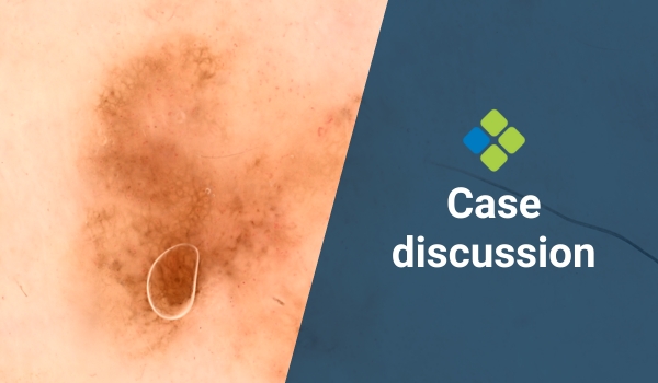
How would you manage this large pigmented lesion?
Case discussion: 20x17mm pigmented lesion on anterior leg of 70-year-old man. Growing over years, with new raised area centrally. What do you think?
Case discussion: 20x17mm pigmented lesion on anterior leg of 70-year-old man. Growing over years, with new raised area centrally. What do you think?

Read more about how to manage athletes with acute musculoskeletal injuries acquired from sport, including treatment protocols and psychological support.

Prof David Wilkinson looks at a recent paper on the treatment of locally advanced and metastatic basosquamous carcinoma.

In this webinar in collaboration with La Roche-Posay, Dr Monisha Gupta covers how to identify and treat pigmentary disorders in primary care.
Case discussion: 5x4mm flat pigmented lesion found on posterior right leg of 65-year-old female. What do you think?

Read more about how GPs can recognise the signs of domestic violence, feel confident asking about it, and provide appropriate support for at-risk women.