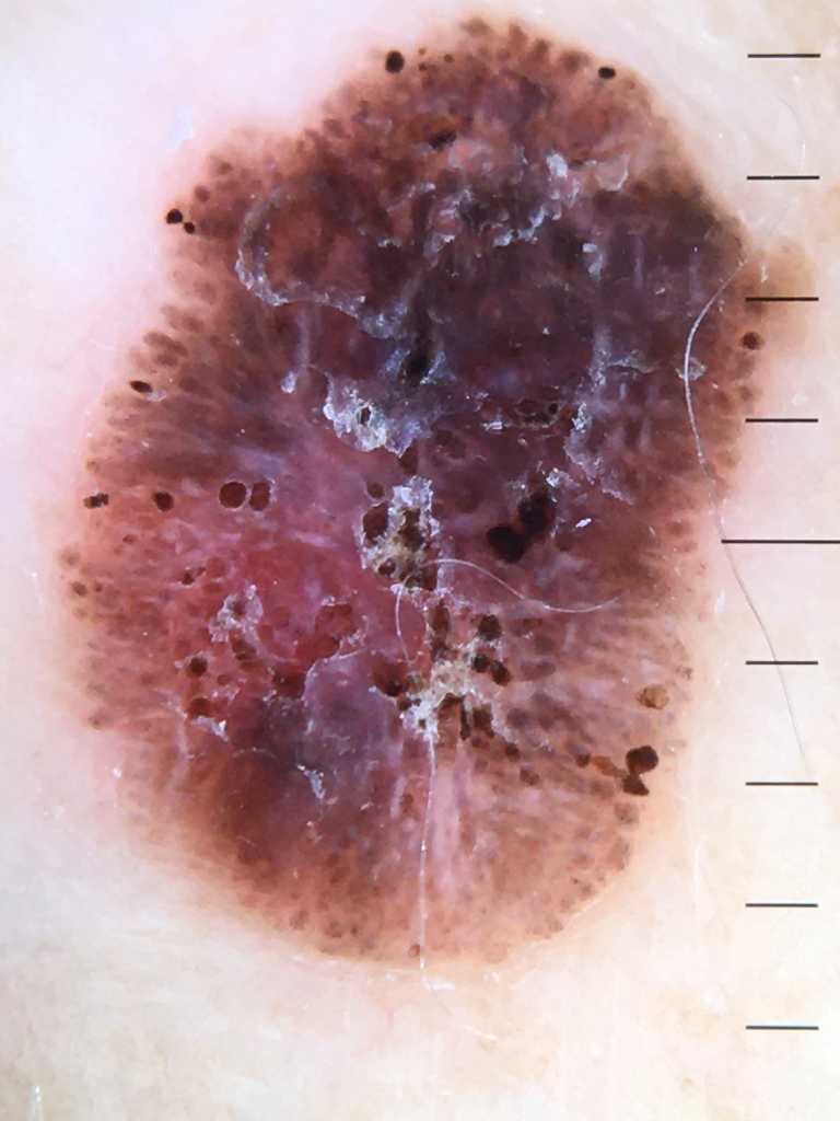
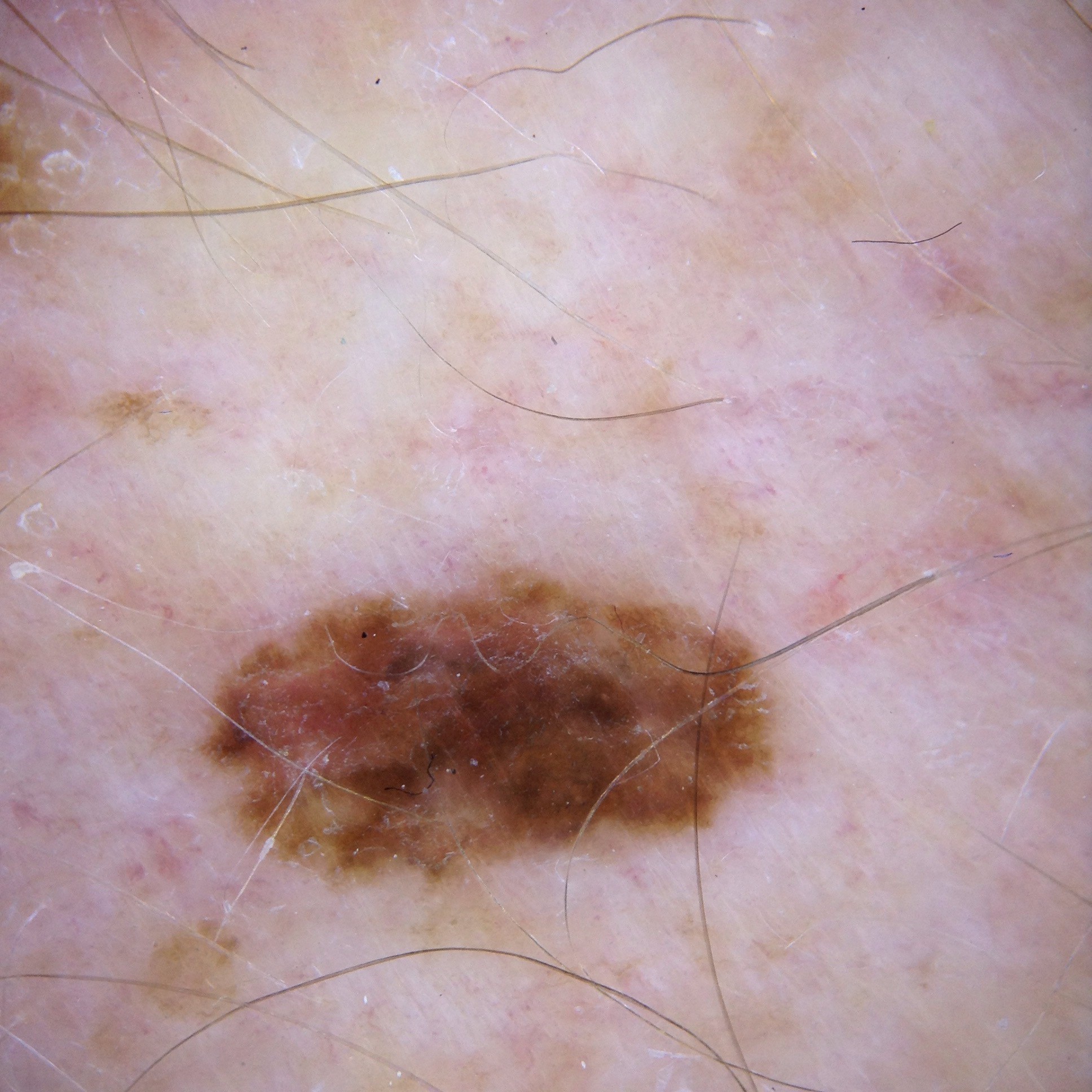
#=$visible?>
Case discussion: How would you treat this patient? [17 October]
In this week’s case discussion, submitted by Dr Magdy Malek, we look at a 73-year-old male patient who presented for a full-body skin cancer check and this pigmented lesion was found on his lower back. The patient has a history of multiple melanomas in the past.
What do you think of the dermoscopy, and what would you do?
Update
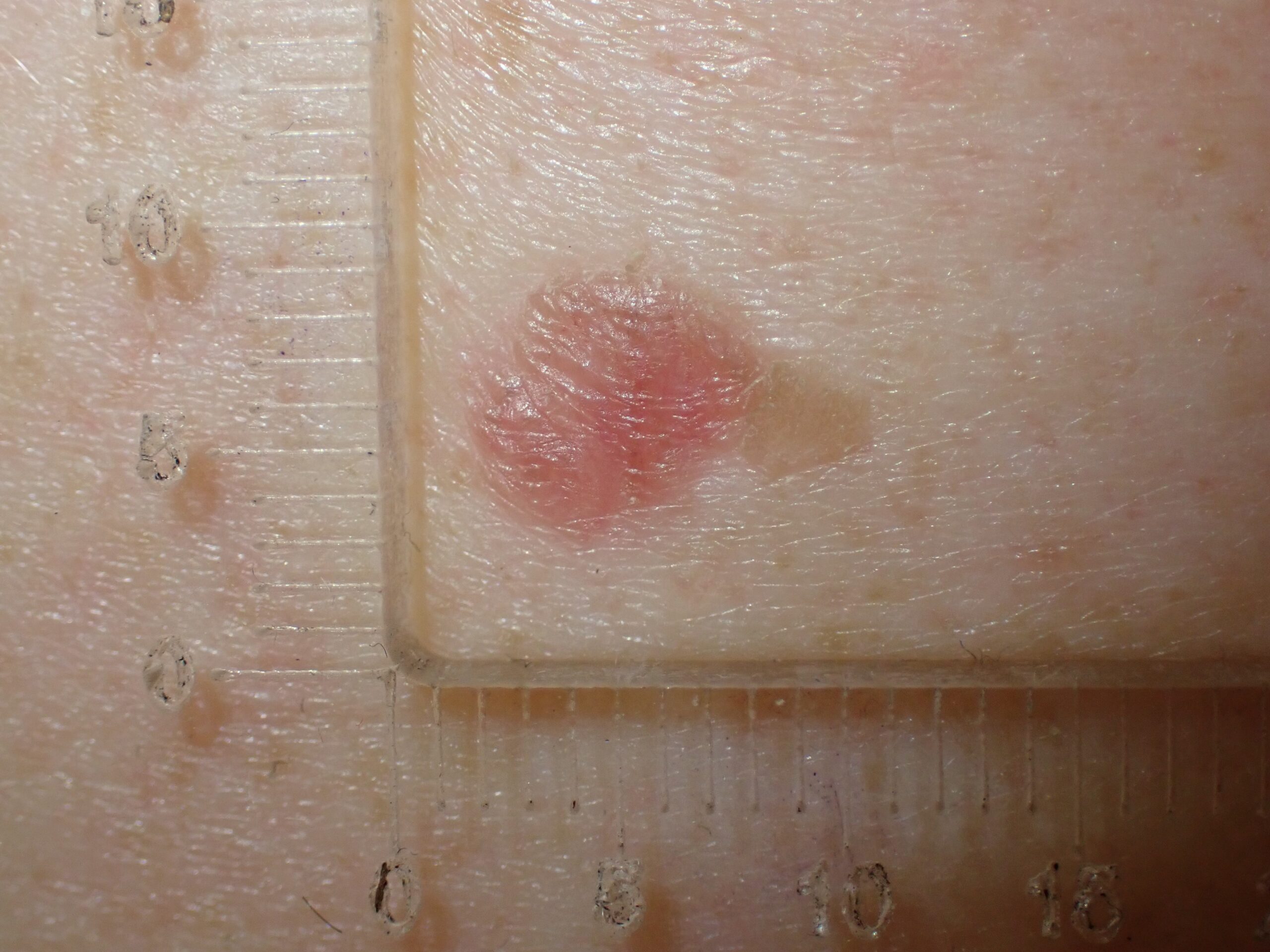
The lesion was shaved and histopathology revealed a Clark 2 melanoma with Breslow thickness 0.4mm. What next?
Would you like to obtain advice or share your experience with your colleagues and Prof David Wilkinson in the weekly blog case discussion?
Participate with your cases so that we can learn together!
Submit your case here or send details to [email protected]
Leave a Reply Cancel reply
16 comments on “Case discussion: How would you treat this patient? [17 October]”
Subscribe
Receive the newest case studies, free video tutorials and research articles right in your inbox.
All features of malignant melanoma..!
Remove asap
Melanoma. Excise
Strong history of Melanoma, This new lesion has asymmetrical colour, with areas of regression and deranged networking and pigmentation. I believe this is a Malignant Melanoma and needs excision biopsy first , viewing for wider excision later depends on the depth.
Different shades and structure with dark blotch. Ugly duckling. Brown snd red must be taken serious. Excise it. DD; mm
Superficial spreading melanoma
Wide excision biopsy is safe option
ASYMMETRICAL LESION WITH AREAS OF LIGHT AND DARK BROWN STRUCTURELESS, THERE IS A SHINY WHITE AREA AT 9 CLOCK, THERE ARE ONE OR TWO PERIPHERAL STREAKS AT 3 CLOCK
MELANOMA FOR EXCISION WITH SAFETY MARGIN
This is clinically a melanoma with depth so fully excise with 2 mm margins and await the depth to determine the margin for clearance ie 10- 20 mm or if SNB be required.
Melanoma? Excision.
polygons, multiple colours, regression. shave the lesion, likely SSM
Clinically a high-risk patient. A new lesion on his lower back?
Dermatoscopically = Chaos: atypical; structure, colour and boarder. (a polaroid image would have been better)
Atypical network, some irregular peripheral streaks. Irregular blotches, with many shades of brown. An erythematous area without blood vessels clearly seen.
IMPRESSION= a superficial spreading melanoma until proved otherwise. The differential diagnoses would be an atypical lentiginous junctional naevus vs an irritated reticulated seborrhoeic keratosis.
PLAN = a full excision with 2mm margins. Further management would be based on histology.
The lesson is suspicious. It has iborder rregularity, areas of regression, , different colour and density.
I will excise the lesion.
The history almost enough to suggest ‘remove this lesion’–but has features of irregular pigmented clumping /assymetry to suggest another melanoma –wide excision
Suspicious pigmented 3/3
Excision biopsy 2mm margins
FURTHER MANAGEMENT= A re excision with a 5mm margin + deep down to the fascia. 3 monthly skin checks for 1 year then 6 monthly for life
Full re- excision with 10mm margin and down to fascia. Confirm nil change in classification that may need more aggressive management.
Review 6 mnths and then annual skin checks.
Suggest full body mapping.