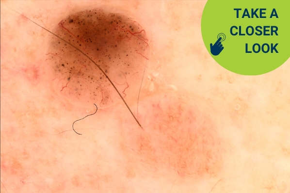
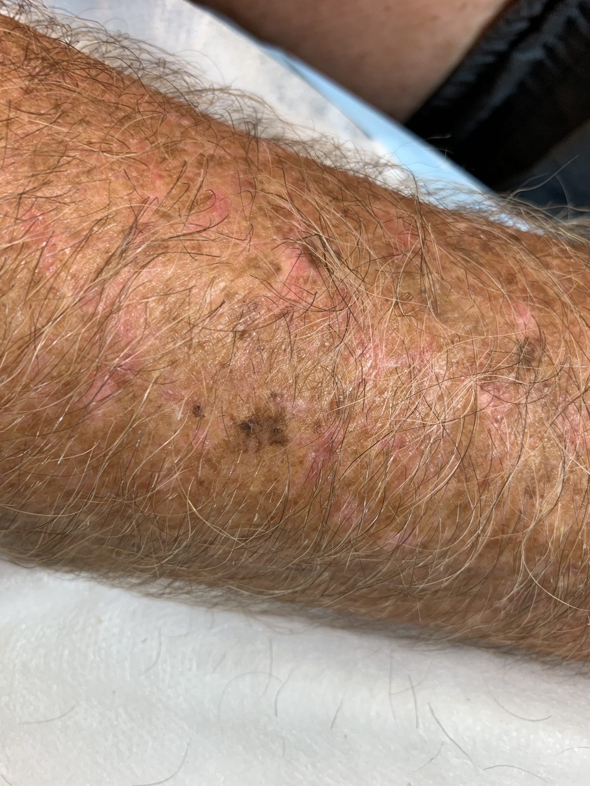
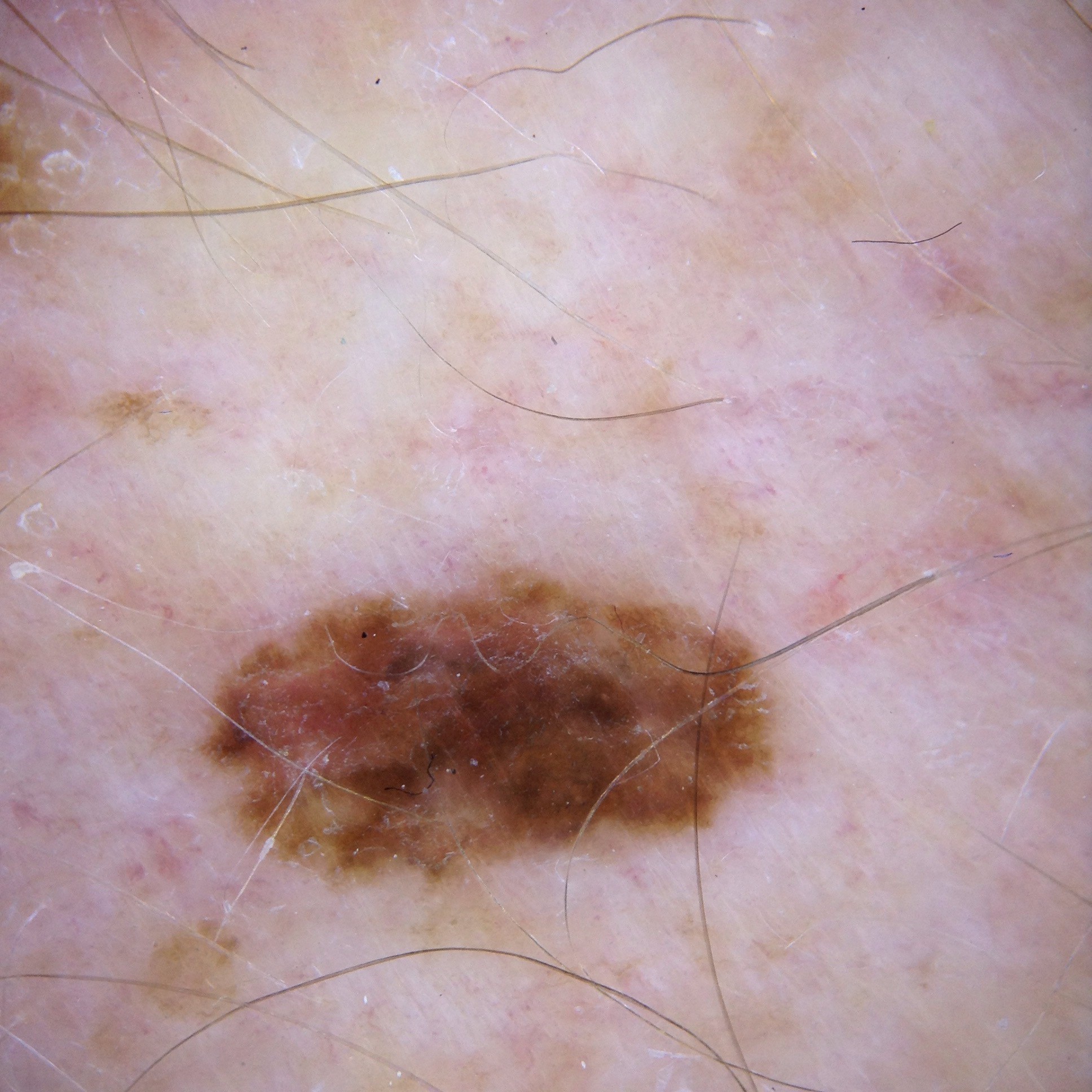
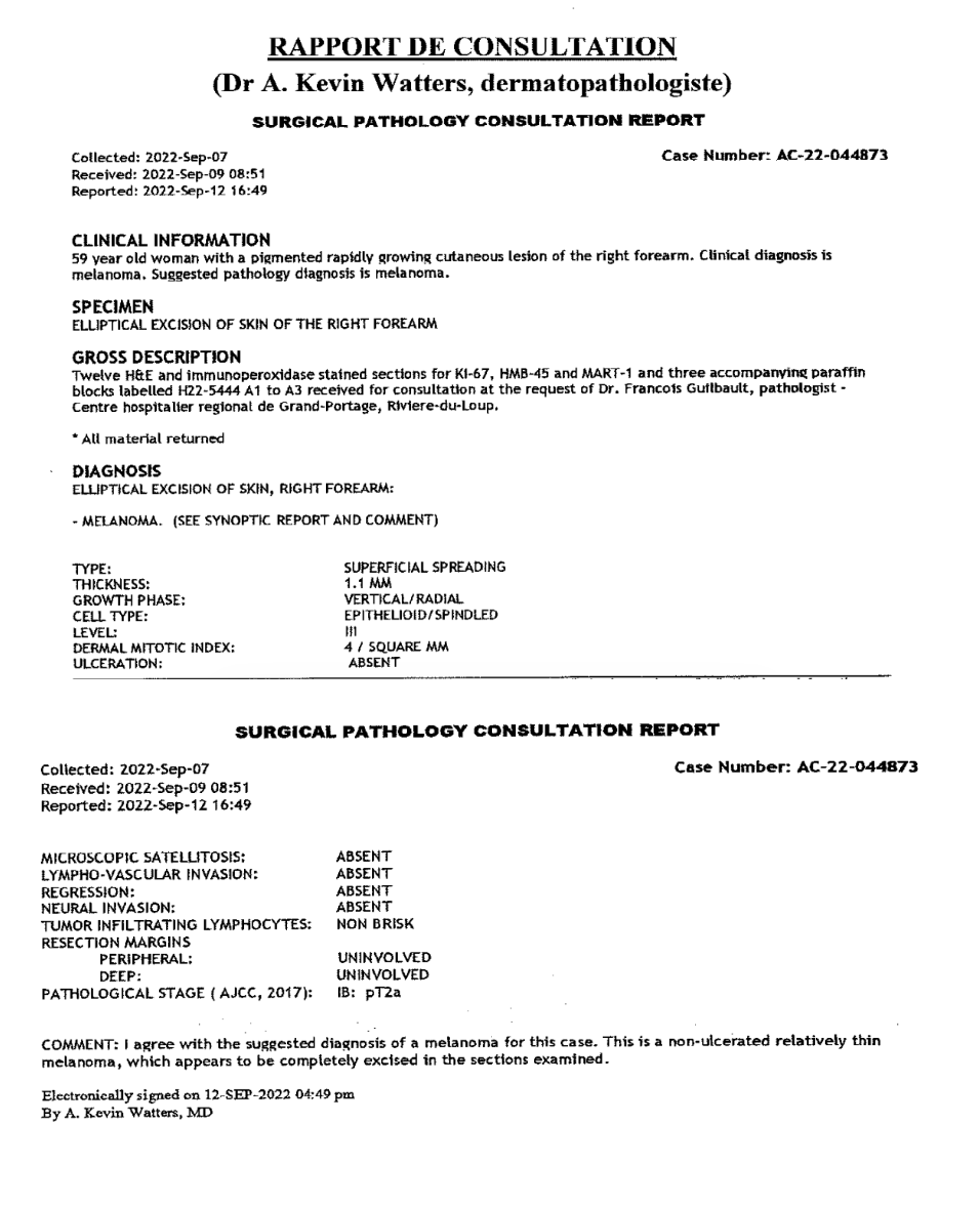
#=$visible?>
Case discussion: How would you treat this patient? [3 October]
Posted on by Abbie Shortt
This week’s case discussion from Dr Céline Bordeleau features a 59-year-old female patient with no previous skin cancers, who presented with a lesion on her right arm that had been present for two years and slowly growing.
What do you make of the dermoscopy?
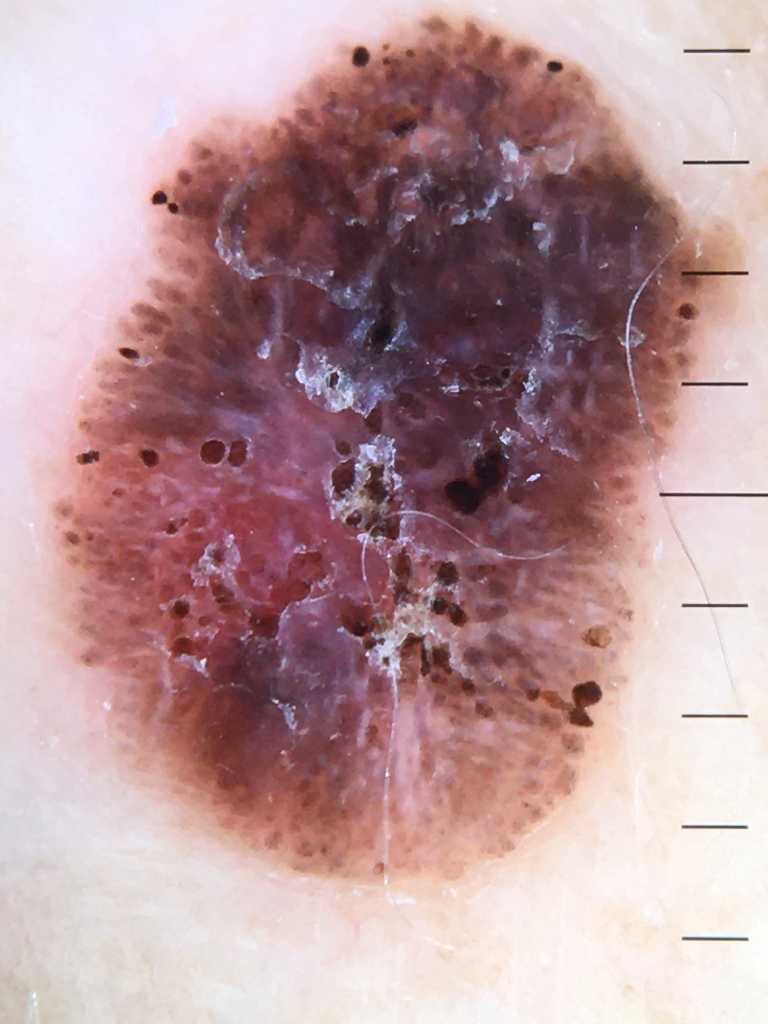
Update:
Here is the pathology. What next?
Would you like to obtain advice or share your experience with your colleagues and Prof David Wilkinson in the weekly blog case discussion?
Participate with your cases so that we can learn together!
Submit your case here or send details to [email protected]
Leave a Reply Cancel reply
17 comments on “Case discussion: How would you treat this patient? [3 October]”
Subscribe
Receive the newest case studies, free video tutorials and research articles right in your inbox.
This definitely needs excision biopsy with 2 mm margins.
I think the dermoscopy shows loss of architecture and pseudopods on the margins , asymmetry and white lines.
Looks like a spitzoid melanoma with white lines on polarisation, peripheral pseudopods, excision biopsy or shave of the lesion en bloc
This is a clear melanoma case. Chaos all the way. Wide excision with a margin of 1cm.
Although there’s a decent chance of Seb k, the chaos, pseudopods, white lines and growth (+/- patient concern) warrant excision.
Black and red clods multiple. Ulcerated lesion. White lines. Most likely a BCC but DDX is Melanoma
To me, there are mixed features.
The history and overall appearance suggest a pigmented seborrheic keratosis to me.
On the other hand the dermoscopy shows some pseudopod formation, variegated pigment, clump formation, and some veiling.
I would biopsy the lesion to resolve any doubt.
Pigmented lesion no network seen.
Pseudopods present.
Assymetric colour
Blue-grey areas suggesting regression
Scale
Suspicious with D/D SebK
Excision biopsy 2mm margins
It looks like a long-standing Seb. k with concerning signs of Melanoma .
I would shave it or excise it with 2 mm margin .
Multiple clods with different sizes and colours/ with pseudopods at periphery. An ulcerated area? White lines. BCC vs spitizoid melanoma.
Clinically = a new solitary growing pigmented lesion on a 59-year-old= a red flag for concern.
Dermatoscopically= A pigmented multicomponent globular/structureless lesion. Clues for a melanoma =Grey blue structureless areas; white lines, peripheral black globules + variable globules as well as irregular streaks. An atypical spitzoid naevus could look like this.
Of note there is central haemorrhagic spots.
Clues for an irritated seb. K.= crypts? (non-polarized view would have been good), fat fingers and acanthotic areas
IMPRESSION = A MELANOMA UNTIL PROVED OTHERWISE, but could be a Spitzoid Naevus vs an Irritated Seb K
PLAN = A full excision with 2mm margin, and further management based on histology
an asymmetrical brown lesion, atypical clods, shiny white areas and streaks, pseudo pods, melanoma excision with safety margin
Atypical lesion with pseudopods in periphery and Clods distributed asymmetrically within the lesion.
Ulceration 8 o’clock? White lines in the center.
Excision probably MM
Malignant melanoma on Rt arm
asymmetrical multiple colours peripheral black clods at periphery white lines
could be a melanoma
either shave or excision biopsy
chaotic pattern with clue – pseudopod at peripheral , whiteline on polarised view
Likely Invasive Melanoma
Will do exicisional biopsy 2 mm margin up to mid fat layer .
_Other DDX could be pigmented Seb keratosis though .
Histology = Superficial Invasive melanoma, Breslow 1.1 mitotic index 4 1B:pT2a
Management would be a referral for a wider excision and a discussion re the benefits/risks of a SLNB .
Again a late comment. Black blue grey and red are a red flag for malignancy and with those black clods scattered thru and irregularly on periphery it can only be a spizoid melanoma. Added the areas of regression and white crystalline lines adds to melanoma. Bcc has blue-black clods. Re seb k do a tape test if think could be superficial blood or keratin and then do dermoscopy again.
The arm has sufficient skin to do full initial 5mm elliptical style excision. From there further management can be decided after histology known.