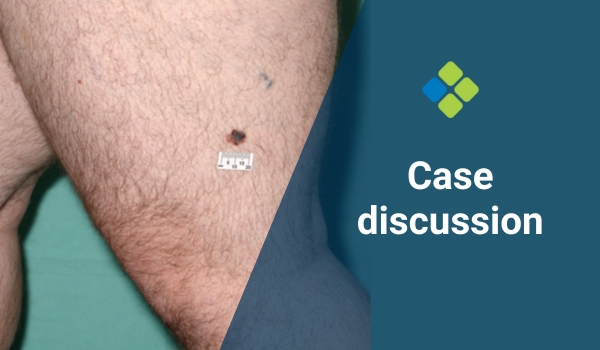
Case discussion: How would you treat this patient? [18 July]
Posted on by Abbie Shortt
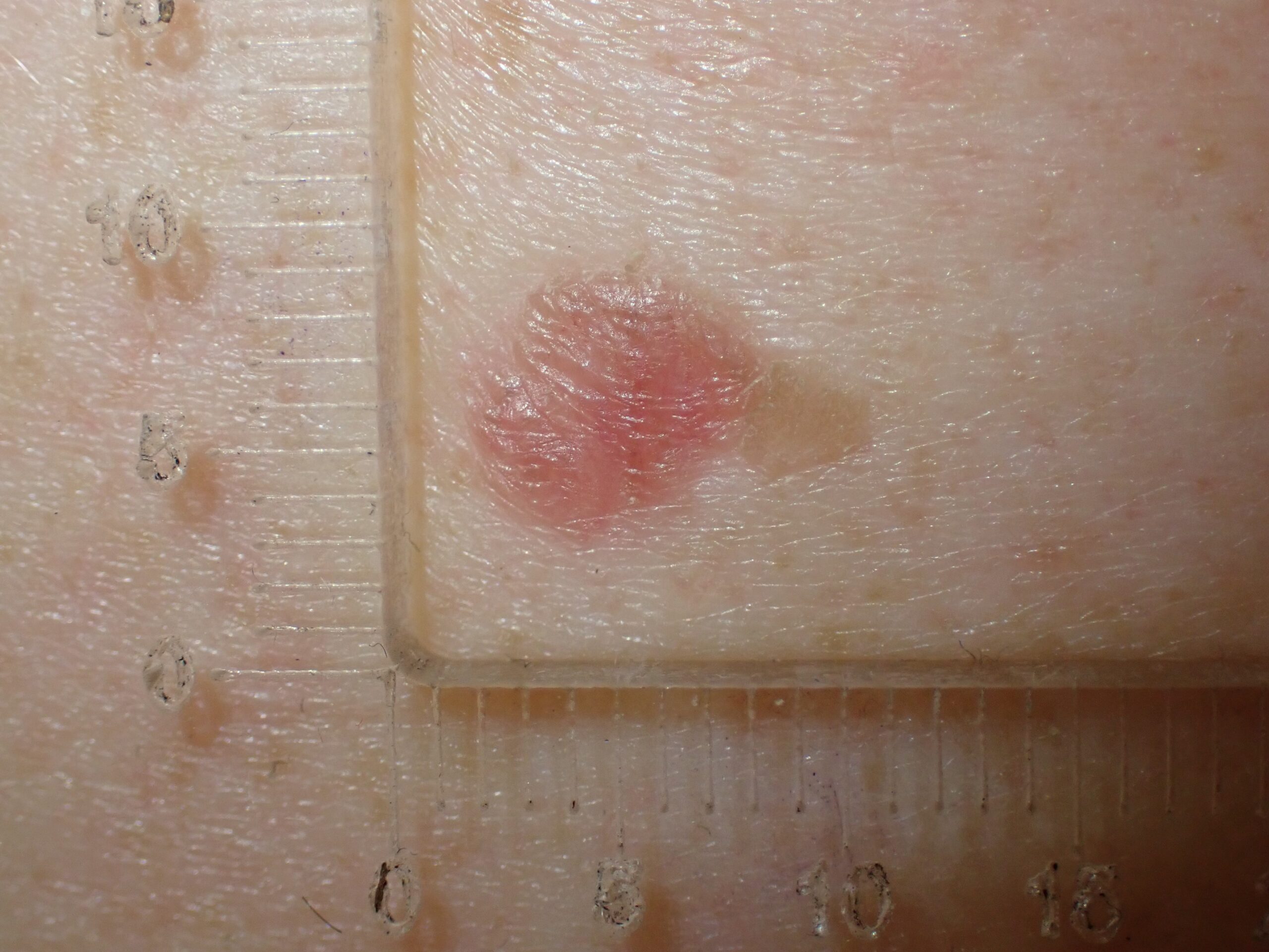
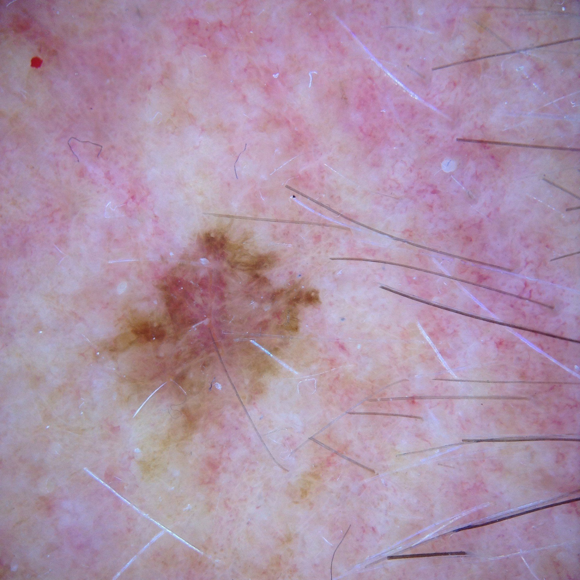
This week, we discuss an interesting case from Dr Tim Aung featuring an asymptomatic 70-year-old male who presented for a total body skin examination. A small pink patch was noted – see below images.
What do you think? What would you do?
Update
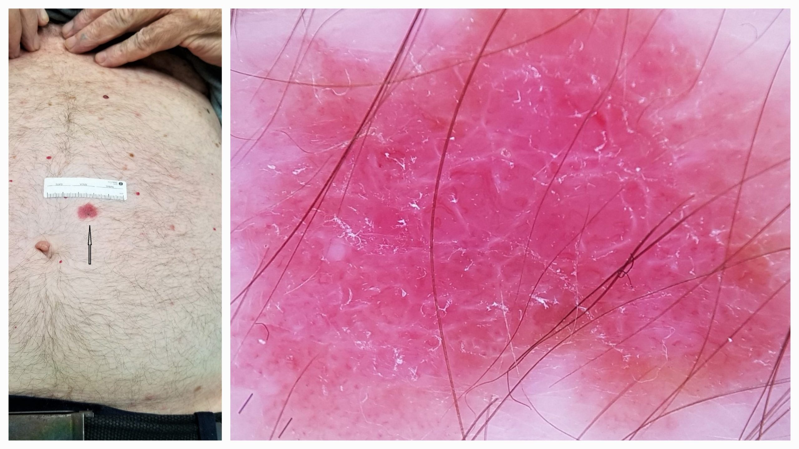
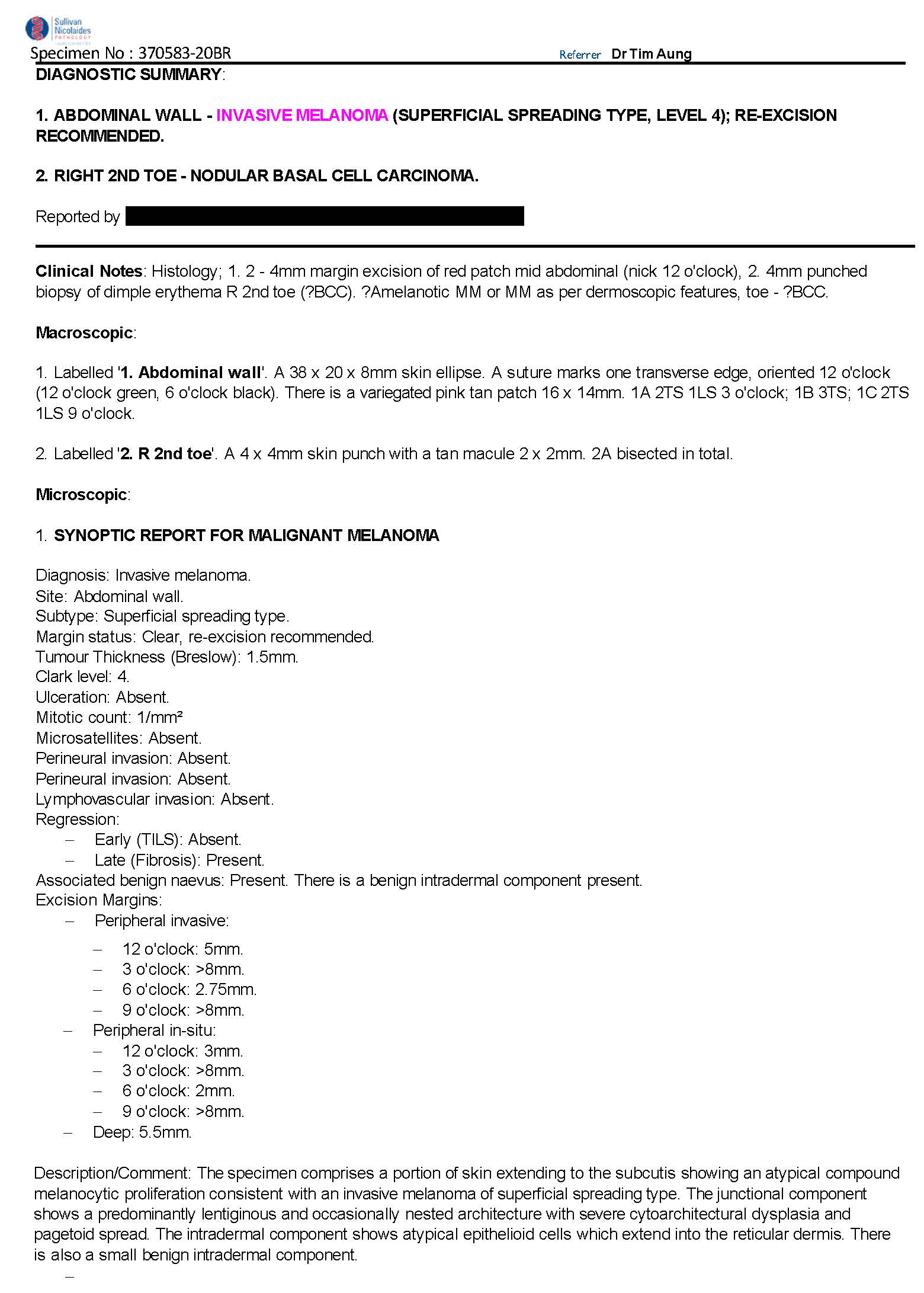
Here is the pathology report. What next?
– Prof David Wilkinson
We encourage you to participate in the case discussions and submit your own clinical images and questions, so we can all learn together.
Leave a Reply Cancel reply
27 comments on “Case discussion: How would you treat this patient? [18 July]”
Subscribe
Receive the newest case studies, free video tutorials and research articles right in your inbox.
Biopsy
I’ll take punch bx as there is polymophic blood vessel’s and white lines
History important; was it there previous, is it evolving. Is it painful, does it bleed
Examination; what does it feel like, is it raised or flat, compare to prior examinations
Dermoscopy; small dot vessels at inferior margin – ? bowens, but white line structures
– Without the clinical information to back up would consider this most likely bowens but would need history and exam to confer confidence not to biopsy
Invasive melanoma. shiny white lines on polarized light. Bizarre polymorphic blood vessels. Faint tan patches at the periphery.
An ugly duckling
Irregular in shape
Non-nevus, pink lesion with irregular demarcation lines
Possible SCC
Punch biopsy 6mm
I’d think of BCC or amelanotic melanoma.
On gross exam, Outstanding lesion .. not scabby to think of SCC as a 1st DD.
On dermoscopy, white lines mostly perpendicular. Polymorphic blood vessels.
If they are polarizing specific white lines, likely amelanotic melanoma.
I’d do full excision biopsy rather than a punch not to miss amelanotic melanoma.
I would biopsy to rule out amelanotic melanoma
Amelanotic melanoma
Excision biopsy
?? Melanoma most likely. Amelanotic.
DDX BCC
Pink skin lesion in old age should be considered seriously. I can see some red dots/ polymorphic vessels and not sure if I can call those short curved lines as white lines. In any case this should be taken off to explore the possibility for malignancy like AMM.
Flat BCC very high likely. Will need punch biopsy.
The mass is irregular in shape , though no network is seen and no blue white colour.
I will biopsy it because of the age of the px. And because it’s the only lesion that looks different from the other lesions.
Might be amelanotic melanoma.
Biopsy to rule out amelanotic melanoma.
Excisionsl biopsy 2mm margin
Exclude amelanotic melanoma vs
Regressing melanoma
White lines polymorphic vessels
? Peripheral melanocytic activity at 5 o’clock
Excision Biopsy with 1mm margin.
superficial BCC likely, can punch biopsy it to get a D/D. treatment depends on that further
? Superficial BCC ? Bowen’s disease .
I would go for 4mm punch biopsy to confirm diagnosis.
Single irregular flat lesion on back with no classical dermoscopy features of s BCC or Bowen’s , psoriasis, inflammatory lesion or eczema..
Does it blanch with pressure? Is it new and or growing.
Need to consider amelanotic melanoma top of DDx list.
Next step excisional biopsy with 2mm margins.
.
It definitely needs a biopsy, preferably excisional. Has polymorphic vessels and chrysalids. Differential diagnosis is amelanotic melanoma or irritated seb k.
Excisuon biopsy 2 mi margin
Dermoscopically a melanoma
Pink amorphous ? Macule(on macro)
-white lies generally with suggestion of polygons – some dot vessels -some brown pigment at 5 o’clock.
? Amel MM -? BCC
Excise with 2mm margin
Pink amorphous ? Macule(on macro)
-white lies generally with suggestion of polygons – some dot vessels -some brown pigment at 5 o’clock.
? Amel MM -? BCC
Excise with 2mm margin
A large solitary red plaque ? slightly raised.
Dermoscopic image = erythematous structureless plaque with some white lines+ polymorphic blood vessels, Of note dotted and linear irregular blood vessels. Some peripheral structureless brown pigmentation.
IMPRESSION= an amelanotic melanoma until proved otherwise , Bowens/SCC is the other possibility. A BCC is less likely re the dotted BVs
PLAN , 2mm margin full excision biopsy if you can . re ? amelanotic melanoma, if one thought it was more likely Bowens a shave excision , but I would favor a full excision.
History is important.
Will Biopsy to R/O Amelanotic Melanoma.
History for how long lesion presenting and any recent change! Do a excisional biopsy with 2mm margin
For more info and educative purpose, you may want to refer to this: N Z Med J. 2022 Jul 1; 135(1557):88-92.
(1) https://pubmed.ncbi.nlm.nih.gov/35772116/
(2) https://journal.nzma.org.nz/journal-articles/a-red-scaly-patch-diagnosed-as-hypomelanotic-melanoma
Cheers
I am surprised that so many colleagues propose to biopsy this with an excision biopsy. Why? The advice / guidelines recommend partial / punch biopsy for pink lesions. To my eye, there is nothing on the dermoscopy of this lesion that differentiates between various possible diagnoses. So, I would have done a 3mm punch biopsy, and the diagnoses of melanoma would have been confirmed, but I would not know the Breslow thickness. So, I would then do a 5mm margin excision (assuming the report did NOT show invasion on my punch biopsy), based on a hope that this would be in situ. If the excision showed invasion then I would do a further, final WLE according to Breslow. Very importantly, we agree to biopsy this lesion before treating it (with cryo, for example).