
 1800 867 1390
1 minute read
Research summary: Treatment of advanced basosquamous carcinoma
Prof David Wilkinson looks at a recent paper on the treatment of locally advanced and metastatic basosquamous carcinoma.
Prof David Wilkinson looks at a recent paper on the treatment of locally advanced and metastatic basosquamous carcinoma.

Case discussion: 20x17mm pigmented lesion on anterior leg of 70-year-old man. Growing over years, with new raised area centrally. What do you think?

In this webinar in collaboration with La Roche-Posay, Dr Monisha Gupta covers how to identify and treat pigmentary disorders in primary care.
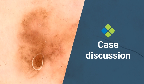
Case discussion: 5x4mm flat pigmented lesion found on posterior right leg of 65-year-old female. What do you think?

Read more about how GPs can recognise the signs of domestic violence, feel confident asking about it, and provide appropriate support for at-risk women.
A discussion on approaches for biopsying suspected melanoma with Prof Cliff Rosendahl and A/Prof Jim Muir at the Skin & Skin Cancer Conference in Brisbane.