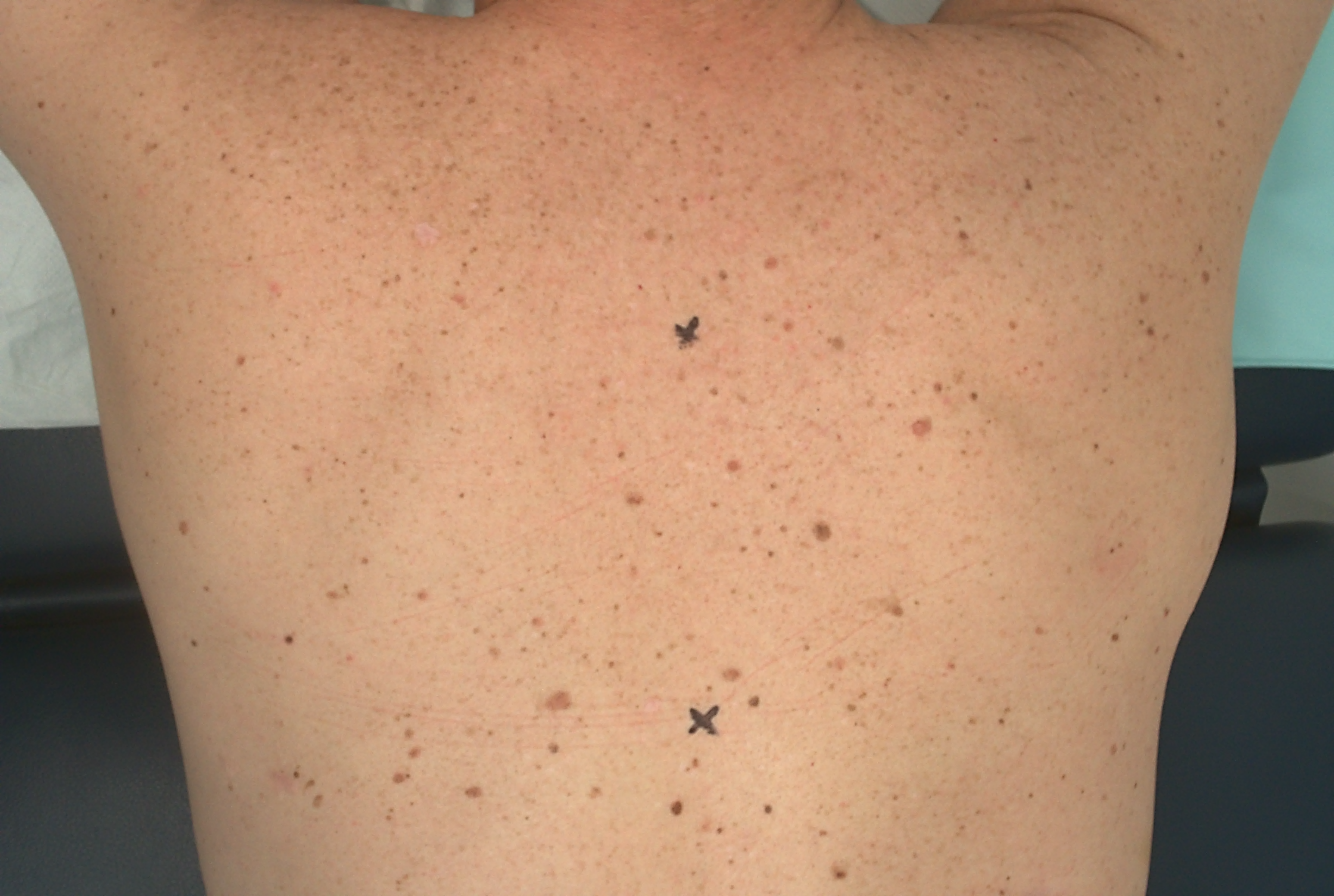
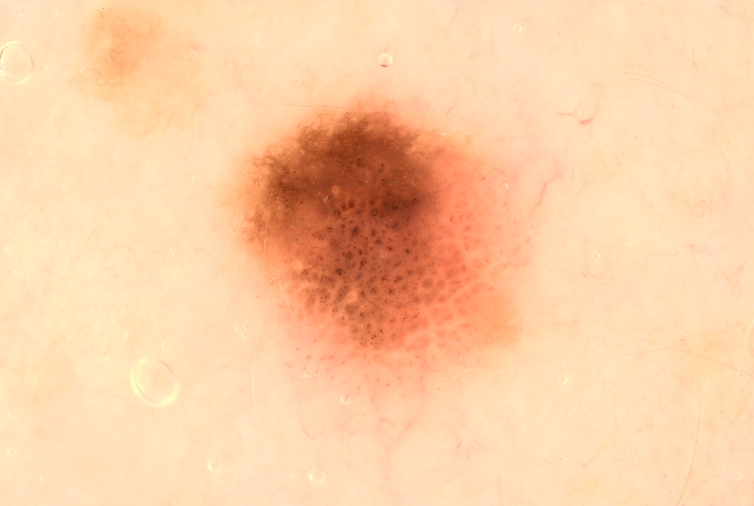
#=$visible?>
Case discussion: How would you treat this patient? [7 September]
Posted on by Abbie Shortt
In this week’s new case discussion from Dr Terry Harvey, we present a patient who came in for a routine skin check:
- 34 year-old male patient
- Asymptomatic lesion on left back
Please review the clinical and dermoscopy images. Any thoughts? What about the pigmented lesion noted? Specific thoughts on the dermoscopy?
Update:
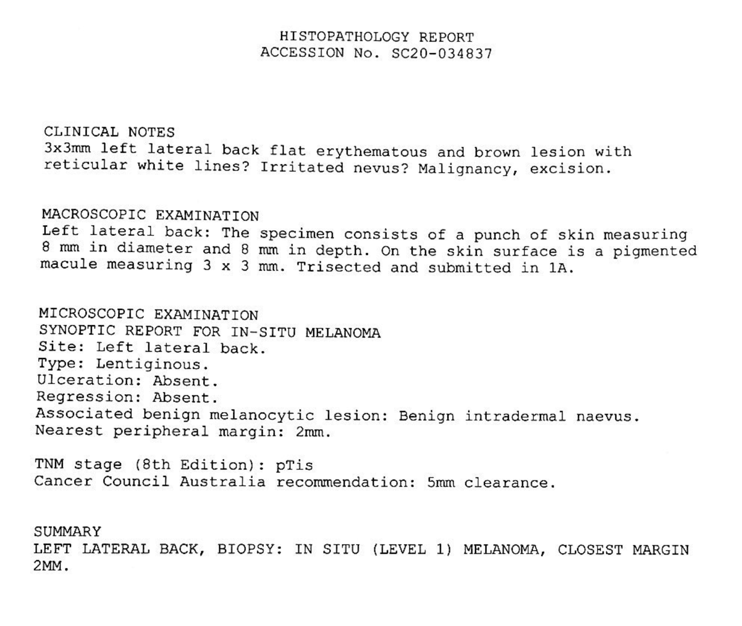
Here is the pathology result. What next?
We encourage you to participate in the case discussions and submit your own clinical images and questions, so we can all learn together.
Learn more about skin cancer medicine in primary care at the next Skin Cancer Certificate Courses:

Leave a Reply Cancel reply
24 comments on “Case discussion: How would you treat this patient? [7 September]”
Subscribe
Receive the newest case studies, free video tutorials and research articles right in your inbox.
Actinic keratosis
early SCC
Asymmetrical etrical with two patterns- in the superior half is a structureless pattern. Excisional biopsy.
Biopsy
no need for biopsy
dermoscope help to make it with 2 step process /peripheral spike and circle pattern clear form SK and it is melanoma
likely,need biopsy to confirm
it seems like a fusion of 2 benign naevus. cobblestone appearance in lower half and regular reticular pattern in upper half.
Benign mole
Chaos and clues, excision biopsy to rule out melanoma
clods and reticular asymmetric lesion with more than 2 colours will excise it with 2mm margin. concerning for melanoma in situ
Melanocytic lesion which has an eccentric dermoscopic island.
In the dermoscopic island, we have clods and globules and some dots.
In the 6 o clock position the lesion is amelanocytic.
Excision Biopsy is recommended as this could be a malignant melanoma in situ.
Dark clods, grey circles, structureless areas and amorphic vessels – ? melanoma – excision biopsy
asymetric structure and asymetric network,
as score 2, I prefer to excise it with 2 mm margin.
sent whole sample to pathology.
Chaos of structure, two patterns – eccentric structureless and brown clods with whitish pseudonetwork. Grey dots, brown lines radial.
Melanoma in situ.
Excision with borders min 5 mm.
Excise with 5 mm margin
Likely melanoma.
Remove with 5 mm margin and wait for Histology to decide what more needs to be done.
? spitzoid melanoma
diagnostic excision with 2 mm margin
Features of chaos and clues indicate the need to perform an excision biopsy ,it would be useful to compare this lesion with the other lesions on the patients back ? Ugly ducking or not .
Benign
Irreg network,irrge outline and areas of dipigmentation .Needs excision biopsy. Person with many moles and sun damaged skin.High risk of developing skin ca .
Excision biopsy
It is difficult to judge with just one clinical and one dermoscopic photo without reference to other pigmented lesions. If the globular pattern is only occuring in this lesion, I will have tendency to cut this out or at least offer short term monitoring as this is by definition an ugly duckling lesion.
1. He is obviously of high risk ie >100mole
2.unfortunate dermoscopic picture has no grid measure
3. A globular Naevus with changes to top section of the lesion hence warrants excisional biopsy with 2 mm margin
Chaotic PL with 2 patterns – reticular and clods asymmetricaly distributed; pink and brown colours. Polymorhous and dotted BVs – Melanoma in Situ
I’m a bit late to the discussion but agree with the clinical notes on the path request. This lesion is chaotic and erythematous with a negative/inverse network; there are several vessels peripherally but I can’t discern their morphology from this photo; it is not a seb k or other benign lesion; I also would have done an excisional biopsy with a 2mm margin, and with the path result, do a 5mm WLE
Frightening case! I wish there was a slightly better photo of how lesion looks to naked eye. ON the current photo it looks no different to rest of back so you would only have done a good job if you put dermoscopy on each and every lesion on his back. Curious as to pink lesion’s dermoscopy features just inferior to the lesion in question.
The dermoscopy shows asymmetry and negative network. RHomboid pattern inferiorly. The negative network also looks like white lines at 5 o’clock. There is dotted and comma vessels inferior and to right. All enough to biopsy entire lesion with 2mm margin.